
Page 230 - Anatomy and Physiology of Farm Animals, 8th Edition
P. 230
Physiology of the Nervous System / 215
Na+ Na+ Na+ 3Na+
VetBooks.ir Extracellular Na+ Na+
space
(b)
Cell (a)
membrane
Cytoplasm ADP
K+ K+ ATP
K+
Large, negatively
(c) charged molecules K+ 2K+
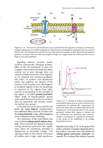
Figure 11-2. The neuron’s cell membrane at rest is polarized by the separation of charges. (a) Positively
charged sodium ions are actively pumped out of the neuron in exchange for potassium ions (in a ratio of
3Na for 3 K). (b) Potassium ions slowly leak out of the neuron in response to their electrical and chemical
gradients. (c) Large molecules with many negative charges are trapped inside the cell because they are too
large to cross the membrane.
Signaling between neurons usually
involves temporarily changing permea
bility of the cell membrane to ions. For Action potential
example, when protein channels that allow
sodium ion to pass through them are gNa
opened, sodium enters the cell in response
to its chemical and electrical gradients;
this influx of positive ions depolarizes Voltage/permeability
(make less negative) the interior of the gK
neuron. In the receptive zone, this happens
in localized regions of the cell membrane
in response to the signals from other
neurons in proportion to the strength of Resting potential
the signal, a so‐called graded potential.
These kinds of depolarizations spread 1 ms
only a small distance from the site at which
they are generated, and become weaker Figure 11-3. Change in sodium and potassium
the further they spread. permeability during neuron action potential. gNa,
conductance for sodium; gK, conductance for
At the junction of the axon with the cell potassium. Early influx of sodium (gNa) drives the
body, the axon hillock, depolarization steepest part of the depolarization of the action
has a different effect, however. It is here that potential. Sodium flow begins to decline and
the action potential, a large, self‐propagating the conductance for potassium (gK) increases.
wave of depolarization, is initiated. Outflow of potassium is a significant contributor
The membrane of the axon hillock and to the return of the membrane to its resting,
axon contains voltage‐gated sodium chan- hyperpolarized state.
nels. These channels are closed at normal
resting membrane potentials but rapidly depolarization (Fig. 11‐3). At the peak of the
open when the membrane potential is action potential (maximal depolarization),
depolarized to threshold voltage. This the sodium channels close and additional
opening results in the inward movement voltage‐gated potassium channels are
of sodium and a large, rapid membrane opened. The closing of the sodium channels