
Page 497 - Anatomy and Physiology of Farm Animals, 8th Edition
P. 497
482 / Anatomy and Physiology of Farm Animals
the transition from anestrus to estrus. begin to multiply under the influence of
Follicular development during the transi-
VetBooks.ir tion to the onset of estrus is characterized LH and form a corpus luteum, or yellow
body. The granulosa cells also continue to
undergo luteinization. Most luteal cells are
by FSH‐driven waves of follicular growth
and the ovaries of these animals will often derived from granulosa cells, but some
have numerous small‐ to medium‐sized cells in the corpus luteum are derived from
follicles capable of steroid hormone syn- the theca interna. Luteal cells are described
thesis. As a result of estrogen production as small and large luteal cells histologically
by these transitional follicles, a female in (Fig. 27‐6).
transition may exhibit estrus‐like behavior, Although a mature follicle and a fully
but is not yet fertile as there is no ovula- formed corpus luteum are about the same
tion. When photoperiod is once again not size, they can be differentiated by sight or
conducive for reproductive behavior, a sec- palpation. The follicle is a sac filled with
ond transition from estrus to anestrus will fluid that has the appearance and feel of a
occur, which can again be associated with blister, while the corpus luteum looks and
nonfertile, estrus‐like behavior. feels solid (Fig. 27‐7).
Blood progesterone levels increase as
corpora lutea grow and develop after
Corpus Luteum ovulation (Fig. 27‐4). When corpora lutea
are fully developed, progesterone secretion
The ovarian corpus luteum (pl. corpora is maximal and plasma levels stabilize.
lutea) is a temporary endocrine organ If fertilization of the ova does not occur
with progesterone as its primary secretory and a pregnancy is not established, the
product. A corpus luteum forms at the corpora lutea spontaneously regress, with
site of each ovulated follicle (Fig. 27‐1), so a relatively rapid decrease in plasma pro-
litter‐bearing animals may have multiple gesterone (Fig. 27‐4). Corpus luteum
corpora lutea on an individual ovary. regression entails apoptotic death of luteal
Sometimes during ovulation small cells, their removal, and the replacement of
blood vessels rupture, and the cavity of the the corpus luteum with connective tissue
ruptured follicle fills with a blood clot, a forming a corpus albicans. If a pregnancy
corpus hemorrhagicum. Whether or not a is established, maternal recognition of
corpus hemorrhagicum forms, the granu- pregnancy occurs, and regression of the
losa cells lining the empty follicular cavity corpus luteum is prevented. This process
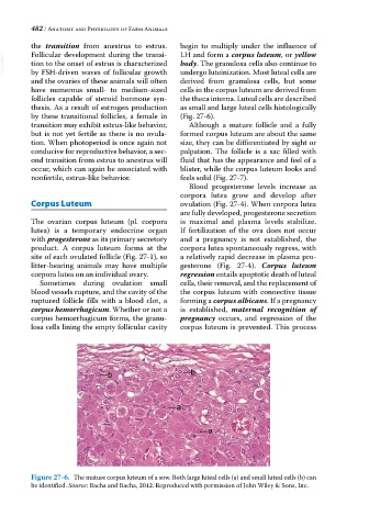
Figure 27-6. The mature corpus luteum of a sow. Both large luteal cells (a) and small luteal cells (b) can
be identified. Source: Bacha and Bacha, 2012. Reproduced with permission of John Wiley & Sons, Inc.