
Page 1001 - Veterinary Immunology, 10th Edition
P. 1001
Atopic dogs commonly present with pruritus. Initially there may be
VetBooks.ir no obvious skin lesions, but this progresses to diffuse erythema
(Table 30.2). Animals may present with the allergic triad: face
rubbing, axillary pruritus, and foot licking. Lesions occur most
commonly on front feet, the ventral abdomen, and in the inguinal
and axillary regions, although they may be found anywhere on the
body. These lesions are secondary to the intense pruritus and vary
from acute erythema and edema to chronic secondary changes
including crusting, scaling, hyperpigmentation, lichenification, and
pyoderma. Some animals also develop rhinitis, otitis externa, or
conjunctivitis. Dogs may also develop focal “hot spots” or urticaria
(Fig. 30.6). Secondary bacterial or yeast infections complicate the
disease. Depending on the inducing allergen, the extrinsic disease
may or may not be seasonal and relapsing. Once it starts, it tends to
get progressively more severe unless treated.
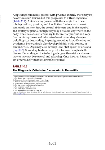
TABLE 30.2
The Diagnostic Criteria for Canine Atopic Dermatitis
The International Task Force on Canine Atopic Dermatitis has listed eight diagnostic criteria for this disease:
1. The disease mainly occurs in indoor dogs.
2. Disease onset occurs in animals under 3 years of age.
3. Animals develop a corticosteroid-responsive pruritus.
4. This pruritus is initially without obvious lesions.
5. The pruritus eventually affects the front feet.
6. The ear pinnae are affected next.
7. The ear margins are affected next.
8. The dorsolumbar area remains unaffected.
Any combination of five criteria from this set will diagnose atopic dermatitis with a sensitivity of 85% and a specificity of
79% (Chapter 42).
1001