
Page 1094 - Veterinary Immunology, 10th Edition
P. 1094
VetBooks.ir
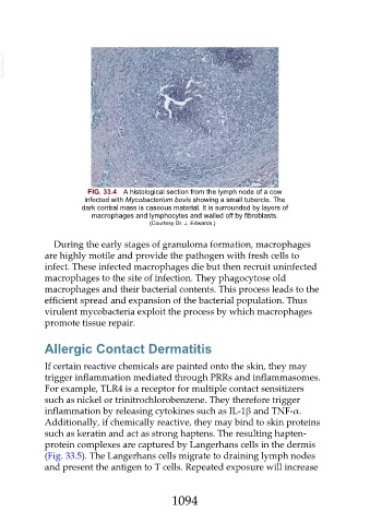
FIG. 33.4 A histological section from the lymph node of a cow
infected with Mycobacterium bovis showing a small tubercle. The
dark central mass is caseous material. It is surrounded by layers of
macrophages and lymphocytes and walled off by fibroblasts.
(Courtesy Dr. J. Edwards.)
During the early stages of granuloma formation, macrophages
are highly motile and provide the pathogen with fresh cells to
infect. These infected macrophages die but then recruit uninfected
macrophages to the site of infection. They phagocytose old
macrophages and their bacterial contents. This process leads to the
efficient spread and expansion of the bacterial population. Thus
virulent mycobacteria exploit the process by which macrophages
promote tissue repair.
Allergic Contact Dermatitis
If certain reactive chemicals are painted onto the skin, they may
trigger inflammation mediated through PRRs and inflammasomes.
For example, TLR4 is a receptor for multiple contact sensitizers
such as nickel or trinitrochlorobenzene. They therefore trigger
inflammation by releasing cytokines such as IL-1β and TNF-α.
Additionally, if chemically reactive, they may bind to skin proteins
such as keratin and act as strong haptens. The resulting hapten-
protein complexes are captured by Langerhans cells in the dermis
(Fig. 33.5). The Langerhans cells migrate to draining lymph nodes
and present the antigen to T cells. Repeated exposure will increase
1094