
Page 125 - Veterinary Immunology, 10th Edition
P. 125
VetBooks.ir
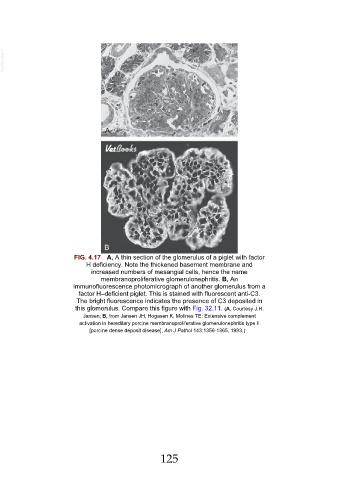
FIG. 4.17 A, A thin section of the glomerulus of a piglet with factor
H deficiency. Note the thickened basement membrane and
increased numbers of mesangial cells, hence the name
membranoproliferative glomerulonephritis. B, An
immunofluorescence photomicrograph of another glomerulus from a
factor H–deficient piglet. This is stained with fluorescent anti-C3.
The bright fluorescence indicates the presence of C3 deposited in
this glomerulus. Compare this figure with Fig. 32.11. (A, Courtesy J.H.
Jansen; B, from Jansen JH, Hogasen K, Mollnes TE: Extensive complement
activation in hereditary porcine membranoproliferative glomerulonephritis type II
[porcine dense deposit disease], Am J Pathol 143:1356-1365, 1993.)
125