
Page 135 - Veterinary Immunology, 10th Edition
P. 135
VetBooks.ir
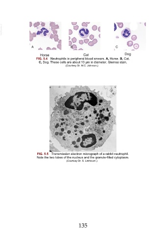
FIG. 5.4 Neutrophils in peripheral blood smears. A, Horse. B, Cat.
C, Dog. These cells are about 10 µm in diameter. Giemsa stain.
(Courtesy Dr. M.C. Johnson.)
FIG. 5.5 Transmission electron micrograph of a rabbit neutrophil.
Note the two lobes of the nucleus and the granule-filled cytoplasm.
(Courtesy Dr. S. Linthicum.)
135