
Page 175 - Veterinary Immunology, 10th Edition
P. 175
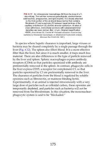
FIG. 6.11  An intravascular macrophage (M) from the lung of a 7-
  VetBooks.ir              siderosomes, phagosomes, and lipid droplets. It is closely attached
                             day-old pig. The cell has numerous pseudopods, electron-dense

                              to the thick portion of the air-blood tissue barrier that contains
                              fibroblasts (F) and a pericyte (P) between basal laminae of the
                             capillary endothelium (E) and the alveolar epithelium. At sites of
                              close adherence, intercellular junctions with subplasmalemmal
                              densities are seen (arrow). Bar = 2 µm. Original magnification
                             ×8000. (From Winkler GC, Cheville NF: Postnatal colonization of porcine lung
                            capillaries by intravascular macrophages: an ultrastructural morphometric analysis,
                                              Microvasc Res 33:224-232, 1987.)


                  In species where hepatic clearance is important, large viruses or

               bacteria may be cleared completely by a single passage through the
               liver (Fig. 6.12). The spleen also filters blood. It is a more effective
               filter than the liver, but since it is much smaller, it traps much less
               material. There are also differences in the type of particle removed

               by the liver and spleen. Splenic macrophages express antibody
               receptors (CD64) so that particles opsonized with antibody are
               preferentially removed in the spleen. In contrast, phagocytic cells in
               the liver express CD35, a receptor for complement C3, so that

               particles opsonized by C3 are preferentially removed in the liver.
               The clearance of particles from the blood is regulated by soluble
               opsonins such as fibronectin, or mannose-binding lectin.
               Experimentally, if an animal is injected intravenously with a very

               large dose of particles such as colloidal carbon, these opsonins are
               temporarily depleted, and particles such as bacteria will not be
               removed from the bloodstream. In this situation, the mononuclear-
               phagocytic system is said to be “blockaded.”


                                                         175