
Page 212 - Veterinary Immunology, 10th Edition
P. 212
VetBooks.ir
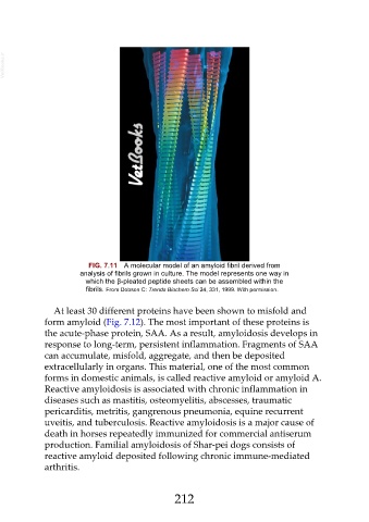
FIG. 7.11 A molecular model of an amyloid fibril derived from
analysis of fibrils grown in culture. The model represents one way in
which the β-pleated peptide sheets can be assembled within the
fibrils. From Dobson C: Trends Biochem Sci 24, 331, 1999. With permission.
At least 30 different proteins have been shown to misfold and
form amyloid (Fig. 7.12). The most important of these proteins is
the acute-phase protein, SAA. As a result, amyloidosis develops in
response to long-term, persistent inflammation. Fragments of SAA
can accumulate, misfold, aggregate, and then be deposited
extracellularly in organs. This material, one of the most common
forms in domestic animals, is called reactive amyloid or amyloid A.
Reactive amyloidosis is associated with chronic inflammation in
diseases such as mastitis, osteomyelitis, abscesses, traumatic
pericarditis, metritis, gangrenous pneumonia, equine recurrent
uveitis, and tuberculosis. Reactive amyloidosis is a major cause of
death in horses repeatedly immunized for commercial antiserum
production. Familial amyloidosis of Shar-pei dogs consists of
reactive amyloid deposited following chronic immune-mediated
arthritis.
212