
Page 276 - Veterinary Immunology, 10th Edition
P. 276
as dendrites (Fig. 10.2). The dendrites increase the cell surface area
VetBooks.ir and thus increase the efficiency of antigen trapping and maximize
contact between DCs and other cell types.
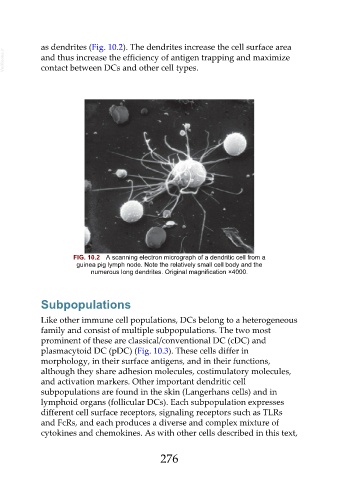
FIG. 10.2 A scanning electron micrograph of a dendritic cell from a
guinea pig lymph node. Note the relatively small cell body and the
numerous long dendrites. Original magnification ×4000.
Subpopulations
Like other immune cell populations, DCs belong to a heterogeneous
family and consist of multiple subpopulations. The two most
prominent of these are classical/conventional DC (cDC) and
plasmacytoid DC (pDC) (Fig. 10.3). These cells differ in
morphology, in their surface antigens, and in their functions,
although they share adhesion molecules, costimulatory molecules,
and activation markers. Other important dendritic cell
subpopulations are found in the skin (Langerhans cells) and in
lymphoid organs (follicular DCs). Each subpopulation expresses
different cell surface receptors, signaling receptors such as TLRs
and FcRs, and each produces a diverse and complex mixture of
cytokines and chemokines. As with other cells described in this text,
276