
Page 309 - Veterinary Immunology, 10th Edition
P. 309
VetBooks.ir
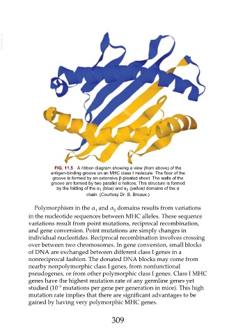
FIG. 11.5 A ribbon diagram showing a view (from above) of the
antigen-binding groove on an MHC class I molecule. The floor of the
groove is formed by an extensive β-pleated sheet. The walls of the
groove are formed by two parallel α helices. This structure is formed
by the folding of the α (blue) and α (yellow) domains of the α
2
1
chain. (Courtesy Dr. B. Breaux.)
Polymorphism in the α and α domains results from variations
1 2
in the nucleotide sequences between MHC alleles. These sequence
variations result from point mutations, reciprocal recombination,
and gene conversion. Point mutations are simply changes in
individual nucleotides. Reciprocal recombination involves crossing
over between two chromosomes. In gene conversion, small blocks
of DNA are exchanged between different class I genes in a
nonreciprocal fashion. The donated DNA blocks may come from
nearby nonpolymorphic class I genes, from nonfunctional
pseudogenes, or from other polymorphic class I genes. Class I MHC
genes have the highest mutation rate of any germline genes yet
−3
studied (10 mutations per gene per generation in mice). This high
mutation rate implies that there are significant advantages to be
gained by having very polymorphic MHC genes.
309