
Page 925 - Veterinary Immunology, 10th Edition
P. 925
VetBooks.ir
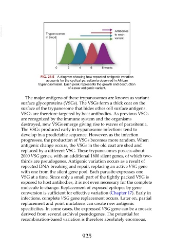
FIG. 28.5 A diagram showing how repeated antigenic variation
accounts for the cyclical parasitemia observed in African
trypanosomiasis. Each peak represents the growth and destruction
of a new antigenic variant.
The major antigens of these trypanosomes are known as variant
surface glycoproteins (VSGs). The VSGs form a thick coat on the
surface of the trypanosome that hides other cell surface antigens.
VSGs are therefore targeted by host antibodies. As previous VSGs
are recognized by the immune system and the organisms
destroyed, new VSGs emerge giving rise to waves of parasitemia.
The VSGs produced early in trypanosome infections tend to
develop in a predictable sequence. However, as the infection
progresses, the production of VSGs becomes more random. When
antigenic change occurs, the VSGs in the old coat are shed and
replaced by a different VSG. These trypanosomes possess about
2000 VSG genes, with an additional 1600 silent genes, of which two-
thirds are pseudogenes. Antigenic variation occurs as a result of
repeated DNA breaking and repair, replacing an active VSG gene
with one from the silent gene pool. Each parasite expresses one
VSG at a time. Since only a small part of the tightly packed VSG is
exposed to host antibodies, it is not even necessary for the complete
molecule to change. Replacement of exposed epitopes by gene
conversion is sufficient for effective variation (Chapter 17). Early in
infections, complete VSG gene replacement occurs. Later on, partial
replacement and point mutations can create new antigenic
specificities. In some cases, the expressed VSG gene can be a mosaic
derived from several archival pseudogenes. The potential for
recombination-based variation is therefore absolutely enormous.
925