
Page 968 - Veterinary Immunology, 10th Edition
P. 968
VetBooks.ir Response of Mast Cells to Antigen
Although there are numerous ways by which mast cells can
degranulate, the best studied of these is mediated by IgE bound to
FcεRI on the cell surface (Fig. 29.8). Mast cells coated in this way are
primed to bind antigen. The mast cell can reside in tissues, with its
attached IgE acting like a mine in a minefield. If an antigen binds to
this IgE, the mast cell will release its granules into the surrounding
tissues.
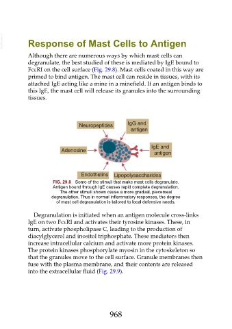
FIG. 29.8 Some of the stimuli that make mast cells degranulate.
Antigen bound through IgE causes rapid complete degranulation.
The other stimuli shown cause a more gradual, piecemeal
degranulation. Thus in normal inflammatory responses, the degree
of mast cell degranulation is tailored to local defensive needs.
Degranulation is initiated when an antigen molecule cross-links
IgE on two FcεRI and activates their tyrosine kinases. These, in
turn, activate phospholipase C, leading to the production of
diacylglycerol and inositol triphosphate. These mediators then
increase intracellular calcium and activate more protein kinases.
The protein kinases phosphorylate myosin in the cytoskeleton so
that the granules move to the cell surface. Granule membranes then
fuse with the plasma membrane, and their contents are released
into the extracellular fluid (Fig. 29.9).
968