
Page 124 - Clinical Manual of Small Animal Endosurgery
P. 124
112 Clinical Manual of Small Animal Endosurgery
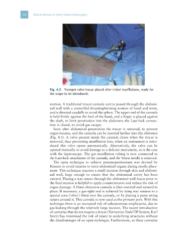
Fig. 4.5 Trumpet valve trocar placed after initial insufflations, ready for
the scope to be introduced.
motion. A traditional trocar-cannula unit is passed through the abdomi-
nal wall with a controlled thrusting/twisting motion of hand and wrist,
and is directed caudally to avoid the spleen. The upper end of the cannula
is held firmly against the heel of the hand, and a finger is placed against
the shaft, to limit penetration into the abdomen; the Luer-lock connec-
tion is closed, to avoid gas escape.
Soon after abdominal penetration the trocar is removed, to prevent
organ trauma, and the cannula can be inserted further into the abdomen
(Fig. 4.5). A valve present inside the cannula closes when the trocar is
removed, thus preventing insufflation loss; when an instrument is intro-
duced this valve opens automatically. Alternatively, the valve can be
opened manually to avoid damage to a delicate instrument, as is the case
with the laparoscope. The gas insufflation tubing is now connected to
the Luer-lock attachment of the cannula, and the Veress needle is removed.
The open technique to achieve pneumoperitoneum was devised by
Hasson to avoid injuries to intra-abdominal organs during needle place-
ment. This technique requires a small incision through skin and abdomi-
nal wall, large enough to ensure that the abdominal cavity has been
entered. Placing a stay suture through the abdominal wall fascia prior to
the final incision is helpful to apply countertension and reduce the risk of
organ damage. A blunt obturator-cannula is then inserted and sutured in
place. If necessary, a gas-tight seal is achieved by tying stay sutures to a
special cone (‘olive’) fitted over the cannula, or by placing a purse-string
suture around it. This cannula is now used as the primary port. With this
technique there is an increased risk of subcutaneous emphysema, due to
gas leaking through the relatively large incision. The recent introduction
of cannulae that do not require a trocar (Ternamian EndoTIP System, Karl
Storz) has minimised the risk of injury to underlying structures without
the disadvantages of an open technique. Furthermore, as these cannulae