
Page 31 - Equine Clinical Medicine, Surgery and Reproduction, 2nd Edition
P. 31
6 CHAPTER 1
VetBooks.ir 1.12 1.13
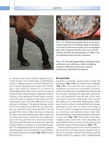
Fig. 1.13 This horse presented with an acute-onset,
moderate lameness of a forelimb, which was localised
to the foot by hoof tester reaction, and a strong digital
pulse. After removal of the shoe, clear areas of fresh
subsolar and white line haemorrhage are visible at the
toe underneath where the shoe was placed.
Fig. 1.12 Note the dropped elbow with flexed carpus
and fetlock seen in this horse, which is exhibiting
temporary radial nerve paresis post a general
anaesthesia in right lateral recumbency.
an obvious reason merits further inspection (e.g. a At exercise
recent wound). The cardinal signs of inflammation Lameness is generally assessed from in front (for
of heat, swelling and pain are noted. Normal reac- forelimbs) (Fig. 1.14) and behind (for hindlimbs)
tion to gentle digital pressure versus that of flinch- (Fig. 1.15) at the walk and trot, in-hand and in a
ing to pain should be assessed. It is common in straight line on a level, hard, even surface. The move-
Thoroughbreds for there to be excessive reaction to ment of the whole horse and individual limbs should
palpation of the distal suspensory ligaments without be evaluated, including foot placement and break-
evidence of any pathology. Careful repeat examina- over, and examination from the side of the horse
tion of the area, or palpation in the same area of the can reveal a shortened stride length and a lowered
contralateral limb, will help differentiate the sig- foot flight arc in a lame limb. Additionally, if lame-
nificance of findings. The ground bearing surface ness is subtle or there is multiple limb involvement,
of the foot is inspected carefully for any abnormali- watching the horse move in circles on the lunge at
ties (e.g. underrun heels) when the limb is off the the trot is useful. A comparison between movement
ground. If the horse is shod, the shoe is inspected on a circle on a soft and hard surface is also help-
for type, wear and nail placement. Hoof testers can ful, particularly in increasing the degree of lameness
be used to assess solar reaction and their application in some cases (Fig. 1.16). Examination of the horse
across the frog and heels may reveal sites of pain. at higher speeds, such as the canter and gallop, and
The shoe should be removed, and the foot inspected when ridden/driven are necessary if the lameness has
further if there is evidence of a foot-related condi- only been noticed in those situations (Fig. 1.17).
tion. The digital pulse to the feet should be palpated. Symmetrical movement is the normal situation in
Bounding pulses are commonly felt in acute lamini- the sound horse and appreciating a change in this is
tis, subsolar abscesses, bruising (Fig. 1.13) and pedal the key to starting to identify the lameness and the
bone fractures. limb(s) involved. In general, with a weight-bearing