
Page 146 - Canine Lameness
P. 146
118 9 JrBay FauBn AacaCoBo cand raahoyBra raoBnhGcyBrao
(A) (B)
(C) (D)
(E) (F)
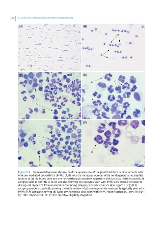
Figure 9.4 Representative examples (A–F) of the appearance of the joint fluid from canine patients with
immune-mediated polyarthritis (IMPA): (A, B) note the increased number of (a) nondegenerate neutrophils
relative to (b) red blood cells and the non-pathologic windrowing pattern that can occur with viscous fluid
samples such as joint fluid; (C, D) samples showing (c) ragocytes seen with IMPA; care should be taken to
distinguish ragocytes from neutrophils containing phagocytized bacteria (see also Figure 9.3C); (D, E)
cytoprep samples clearly illustrating the high number of (a) nondegenerate neutrophils typically seen with
IMPA; (E, F) samples showing (d) lupus erythematous cells seen with IMPA. Magnification: (A): 10×; (B): 50×;
(E): 100× objective; (C, D, F): 100× objective digitally magnified.