
Page 177 - Canine Lameness
P. 177
12.2 Normal Anatomy 149 DISTAL LIMB REGION
(A) (C) (D)
(E)
(B)
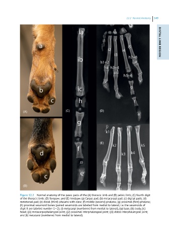
Figure 12.2 Normal anatomy of the paws: pads of the (A) thoracic limb and (B) pelvic limb; (C) fourth digit
of the thoracic limb; (D) forepaw; and (E) hindpaw. (a) Carpal pad; (b) metacarpal pad; (c) digital pads; (d)
metatarsal pad; (e) distal (third) phalanx with claw; (f) middle (second) phalanx; (g) proximal (first) phalanx;
(h) proximal sesamoid bones (paired sesamoids are labeled from medial to lateral; i.e. the sesamoids of
digit II are labeled number 1 + 2); (i) metacarpi (numbered from medial to lateral), (ia) base, (ib) body, (ic)
head; (j1) metacarpophalangeal joint; (j2) proximal interphalangeal joint; (j3) distal interphalangeal joint;
and (k) metatarsi (numbered from medial to lateral).