
Page 54 - Canine Lameness
P. 54
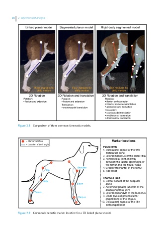
26  2  Objective Gait Analysis


                Linked planar model  Segmented planar model   Rigid-body segmented model


                     Three markers for       Four markers for      Ten markers for
                       stifle motion          stifle motion          stifle motion
                   2D Rotation      2D Rotation and translation  3D Rotation and translation
               Rotation:              Rotation:                  Rotation:
               • flexion and extension  • flexion and extension  • flexion and extension
                                      Translation:               • internal and external rotation
                                      • craniocaudal translation  • abduction and adduction
                                                                 Translation:
                                                                 • craniocaudal translation
                                                                 • mediolateral translation
                                                                 • dorsoventral translation

            Figure 2.8  Comparison of three common kinematic models.


                 = Marker location                                   Marker locations
                 = Location of joint angle
                                                               Pelvic limb
                                                               1. Distolateral aspect of the fifth
                                                                 metatarsal bone
                                                               2. Lateral malleolus of the distal tibia
                                                               3. Femorotibial joint, midway
                                5         6                      between the lateral epicondyle of
                                                                 the femur and the fibular head
                           4                    Shoulder       4. Greater trochanter of the femur
                             Hip               7               5. Iliac crest
                                                               Thoracic limb
                         3
                           Stifle                              6. Dorsal aspect of the scapular
                                            8  Elbow             spine
                                                               7. Acromion/greater tubercle of the
                    2                                            scapulohumeral joint
                      Tarsus                                   8. Lateral epicondyle of the humerus
                                                               9. Ulnar styoloid process/ulnar
                                              9  Carpus          carpal bone of the carpus
                   1
                                              10              10. Distolateral aspect of the fifth
                                                                 metacarpal bone

            Figure 2.9  Common kinematic marker location for a 2D linked planar model.