
Page 175 - Basic Monitoring in Canine and Feline Emergency Patients
P. 175
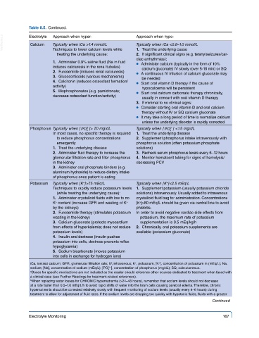
Table 8.5.  Continued.

  VetBooks.ir  Electrolyte  Approach when hyper-         Approach when hypo-
                                                         Typically when iCa <0.8–1.0 mmol/L
                       Typically when iCa >1.4 mmol/L
              Calcium
                       Techniques to lower calcium levels while
                                                           2.  If significant clinical signs (e.g. tetany/seizures/car-
                         treating the underlying cause:    1.  Treat the underlying cause
                                                         diac arrhythmias):
                         1.  Administer 0.9% saline fluid (Na in fluid   ●  Administer calcium (typically in the form of 10%
                       induces calciuresis in the renal tubules)  calcium gluconate) IV slowly (over 5-10 min) or SQ
                         2.  Furosemide (induces renal calciuresis)  ●  A continuous IV infusion of calcium gluconate may
                         3.  Glucocorticoids (various mechanisms)  be needed
                         4.  Calcitonin (reduces osteoclast formation/  ●  Start oral vitamin D therapy if the cause of
                       activity)                            hypocalcemia will be persistent
                         5.  Bisphosphonates (e.g. pamidronate;   ●  Start oral calcium carbonate therapy chronically,
                       decrease osteoclast function/activity)
                                                            usually in concert with oral vitamin D therapy
                                                           3.  If minimal to no clinical signs:
                                                         ●  Consider starting oral vitamin D and oral calcium
                                                            therapy without IV or SQ calcium gluconate
                                                         ●  It may take a long period of time to normalize calcium
                                                            unless the underlying disorder is rapidly corrected
              Phosphorus Typically when [ PO ]> 7.0 mg/dL  Typically when [ PO ] <1.5 mg/dL
                                                                       3-
                                     3-
                                                                       4
                                     4
                       In most cases, no specific therapy is required     1.  Treat the underlying disease
                         to reduce phosphorus concentrations     2.  Supplement phosphorus intake intravenously with
                         emergently                      phosphorus solution (often potassium phosphate
                        1.  Treat the underlying disease  solutions)
                        2.  Administer fluid therapy to increase the      3.  Recheck serum phosphorus levels every 6–12 hours
                       glomerular filtration rate and filter phosphorus    4.  Monitor hematocrit tubing for signs of hemolysis/
                       in the kidney                     decreasing PCV
                        3.  Administer oral phosphate binders (e.g.
                       aluminum hydroxide) to reduce dietary intake
                       of phosphorus once patient is eating
                                                                      +
                                   +
              Potassium  Typically when [K ]>7.5 mEq/L   Typically when [K ]<2.5 mEq/L
                       Techniques to rapidly reduce potassium levels   1.  Supplement potassium (usually potassium chloride
                         (while treating the underlying cause):  solutions) intravenously. Usually added to intravenous
                         1.  Administer crystalloid fluids with low to no    crystalloid fluid bag for administration. Concentrations
                        +
                       K  content (increase GFR and wasting of K     [K ]>80 mEq/L should be given via  central line to avoid
                                                     +
                                                           +
                       by the kidneys)                   phlebitis.
                         2.  Furosemide therapy (stimulates potassium   In order to avoid negative cardiac side effects from
                       wasting in the kidney)              potassium, the maximum rate of potassium
                         3.  Calcium gluconate (protects myocardium    supplementation is 0.5 mEq/kg/h
                       from effects of hyperkalemia; does not reduce    2.  Chronically, oral potassium supplements are
                       potassium levels)                 available (potassium gluconate)
                         4.  Insulin and dextrose (insulin pushes
                       potassium into cells, dextrose prevents reflex
                       hypoglycemia)
                         5.  Sodium bicarbonate (moves potassium
                       into cells in exchange for hydrogen ions)
              iCa, ionized calcium; GFR, glomerular filtration rate; IV; intravenous; K , potassium, [K ], concentration of potassium in (mEq/L); Na,
                                                                   +
                                                         +
              sodium; [Na], concentration of sodium (mEq/L); [PO   ], concentration of phosphorus (mg/dL); SQ, subcutaneous.
                                              3 −
                                              4
              a Doses for specific medications are not included as the reader should reference other sources dedicated to treatment when faced with
              a clinical case (see Further Readings for treatment-related references).
              b When replacing water losses for CHRONIC hypernatremia (>24–48 hours), remember that sodium levels should not decrease
              at a rate faster than 0.5–1.0 mEq/L/h to avoid rapid shifts of water into the brain cells causing cerebral edema. Therefore, chronic
              hypernatremia should be corrected relatively slowly with frequent monitoring of sodium levels (usually every 4–6 hours) during
              treatment to allow for adjustment of fluid rates. If the sodium levels are dropping too quickly with hypotonic fluids, fluids with a greater
                                                                                         Continued
             Electrolyte Monitoring                                                          167