
Page 210 - Basic Monitoring in Canine and Feline Emergency Patients
P. 210
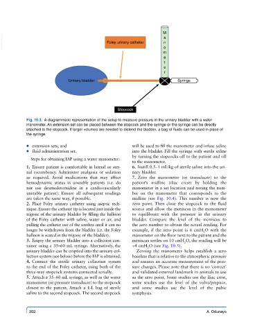
M
  VetBooks.ir                                 Foley urinary catheter  a
                                                                      n
                                                                      o
                                                                      m
                                                                      e
                                                                      t
                                                                      e
                                                                      r
                              Urinary bladder                               Syringe


                                                  Stopcock

            Fig. 10.3.  A diagrammatic representation of the setup to measure pressure in the urinary bladder with a water
            manometer. An extension set can be placed between the stopcock and the syringe or the syringe can be directly
            attached to the stopcock. If larger volumes are needed to distend the bladder, a bag of fluids can be used in place of
            the syringe.


             ● ● extension sets; and                     will be used to fill the manometer and infuse saline
             ● ● fluid administration set.               into the bladder. Fill the syringe with sterile saline
                                                         by turning the stopcocks off to the patient and off
              Steps for obtaining IAP using a water manometer:
                                                         to the manometer.
              1.  Ensure patient is comfortable in lateral or ster-    6.  Instill 0.5–1 mL/kg of sterile saline into the uri-
            nal recumbency. Administer analgesia or sedation   nary bladder.
            as required.  Avoid medications that may affect     7.  Zero the manometer (or transducer) to the
            hemodynamic status in unstable patients (i.e. do   patient’s midline (iliac crest) by holding the
            not  use dexmedetomidine  in a  cardiovascularly   manometer in a set location and noting the num-
            unstable patient). Ensure all subsequent readings   ber on the manometer that corresponds to the
            are taken the same way, if possible.         midline  (see  Fig.  10.4). This  number  is  now  the
              2.  Place  Foley  urinary  catheter  using  aseptic  tech-  zero point. Then close the stopcock to the fluid
            nique. Ensure the catheter tip is located just inside the   source and allow the meniscus in the manometer
            trigone of the urinary bladder by filling the balloon   to equilibrate with the pressure in the urinary
            of the Foley catheter with saline, water or air, and   bladder. Compare the level of the meniscus to
            pulling the catheter out of the urethra until it can no   the zero number to obtain the actual reading. For
            longer be withdrawn from the bladder (i.e. the Foley   example, if the zero point is 6  cmH O with the
                                                                                      2
            balloon is seated in the trigone of the bladder).  manometer on the floor next to the patient and the
              3.  Empty the urinary bladder into a collection con-  meniscus settles on 10 cmH O, the reading will be
                                                                              2
            tainer using a 35–60 mL syringe. Alternatively, the   +4 cmH O (see Fig. 10.5).
                                                               2
            urinary bladder can be emptied into the urinary col-  Zeroing the manometer helps establish a zero
            lection system (see below) before the IAP is obtained.  baseline that is relative to the atmospheric pressure
              4.  Connect the sterile urinary collection system   and ensures an accurate measurement of the pres-
            to the end of the Foley catheter, using both of the   sure changes. Please note that there is no ‘correct’
            three-way stopcock systems connected serially.  and validated external landmark in animals to use
              5.  Attach a 35–60 mL syringe, as well as the water   as the zero point. Some studies use the iliac crest,
            manometer (or pressure transducer) to the stopcock   some studies use the level of the vulva/prepuce,
            closest  to  the  patient. Attach  a  1-L bag  of  sterile   and some studies use the level of the pubic
            saline to the second stopcock. The second stopcock   symphysis.


             202                                                                        A. Odunayo