
Page 1086 - Clinical Small Animal Internal Medicine
P. 1086
1024 Section 9 Infectious Disease
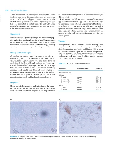
The distribution of Cystoisospora is worldwide. Due to and examined for the presence of characteristic oocysts
VetBooks.ir the fecal–oral route of transmission, cases are associated (Figure 111.1).
It is important to differentiate oocysts of Cystoisospora
with crowded and unhygienic environments. In the
United States, prevalence of canine Cystoisospora spp.
to canine and feline patients. Coprophagy of the feces of
has been estimated to be between 3.1% and 4.4% while spp. from those of Eimeria spp., which are not pathologic
feline Cystoisospora spp. prevalence has been estimated animals such as cattle, sheep, and chickens may lead to
to be between 3.9% and 6.3%. spurious detection of Eimeria spp. in canine and feline
fecal samples. Both Eimeria and Cystoisospora are
species specific and therefore pathogenic only to their
Signalment
target species.
In most surveys, Cystoisospora spp. are detected in pup-
pies and kittens less than 6 months of age. There does not Therapy
appear to be a breed predilection. Patients that are most
susceptible to clinical disease include nursing, recently Asymptomatic adult patients demonstrating fecal
weaned, and immunocompromised dogs and cats. oocysts may be monitored for development of clinical
signs. Patients that meet criteria of history, clinical signs,
and detection of the organism are treated symptomati-
History and Clinical Signs
cally for diarrhea, and concurrently with antiprotozoal
Clinical symptoms are more common in puppies and medications such as ponazuril, sulfonamides, or ampro-
kittens acquired from unsanitary or overcrowded lium (Tables 111.1 and 111.2).
environments. Cystoisospora spp. can cause large or
small bowel diarrhea, although patients may be asymp- Table 111.1 Enteric coccidia of the dog and cat
tomatic despite shedding oocysts. Other clinical symp-
toms reported include anemia, dehydration, vomiting, Organism Diagnostic stage Size (μM)
anorexia, and weight loss. Physical exam findings of
patients with Cystoisospora spp. are nonspecific and can Cystoisospora spp.
include abdominal pain, increased gas or fluid in the Canine
gastrointestinal tract, and thickened loops of bowel. C. canis Oocyst 30 × 38
C. ohioensis Oocyst 19 × 23
C. neorivolta Oocyst 11 × 13
Diagnosis C. burrowsi Oocyst 17 × 20
History, clinical symptoms, and detection of the organ- Feline
ism are needed for a definitive diagnosis of coccidiosis. C. felis Oocyst 30 × 40
20 × 25
Oocyst
C. rivolta
Fecal flotation, centrifugal or passive, can be performed
(a) (b)
Figure 111.1 (a) Sporulated and (b) unsporulated Cystoisospora ohioensis. Source: Courtesy of the National Center for Veterinary
Parasitology, Oklahoma State University.