
Page 1164 - Clinical Small Animal Internal Medicine
P. 1164
1102 Section 10 Renal and Genitourinary Disease
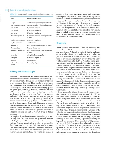
Table 121.1 Select breeds of dogs with familial glomerulopathies ascites, or both, are sometimes noted and consistent
VetBooks.ir Breed Glomerular disease(s) with nephrotic syndrome. Occasionally, there is physical
evidence of thromboembolic disease, such as dyspnea or
Beagle Amyloidosis, primary glomerulopathy a decreased or absent peripheral pulse. Evidence of a
predisposing inflammatory, infectious, or neoplastic
Bernese mountain dog Mesangiocapillary glomerulonephritis process may be detected during the physical examina-
Bull mastiff Glomerulosclerosis tion. The kidneys of affected animals are variable in size.
Bull terrier Hereditary nephritis Animals with chronic kidney disease often have small,
Dalmatian Hereditary nephritis firm, irregularly shaped kidneys, whereas those with less
severe or long‐standing disease often have normal‐sized
Doberman pinscher Glomerulosclerosis, cystic glomerular or, occasionally, enlarged kidneys.
atrophy
English cocker spaniel Hereditary nephritis
English foxhound Amyloidosis Diagnosis
Greyhound Glomerular vasculopathy and necrosis
Newfoundland Glomerulosclerosis When proteinuria is detected, there are three key ele-
Pembroke Welsh corgi Glomerulosclerosis, cystic glomerular ments that need to be assessed: localization, persistence,
atrophy and magnitude. Although proteinuria is the hallmark
Rottweiler Atrophic glomerulopathy of glomerular disease, it can also occur secondary to
Samoyed Hereditary nephritis prerenal, postrenal or renal tubular causes. The magni-
tude of proteinuria is generally assessed via the urine
Shar‐pei Amyloidosis protein:creatinine ratio (UPC). Persistent renal pro-
Soft‐coated wheaten Podocytopathy teinuria that is of high magnitude (i.e., UPC >2) is most
terrier likely of glomerular origin; however, there is no range of
numbers that is diagnostic for any one renal disease and
the overlap in expected ranges is too broad to be clini-
History and Clinical Signs cally reliable. In fact, glomerular lesions have been seen
in dogs without proteinuria. Urine albumin can also
Dogs and cats with glomerular disease can present with be used to assess proteinuria. Microalbuminuria may
a variety of clinical signs, determined by the severity of develop before the UPC is increased. It therefore seems
proteinuria or renal disease and the presence or absence reasonable to conclude that a dog or cat with persistent
of any systemic disorders. They may be asymptomatic, microalbuminuria of increasing magnitude should be
have nonspecific signs (e.g., anorexia, weight loss, lethargy) assessed as having an injurious process to the glomerular
or have signs of more advanced renal disease (e.g., polyu- filtration barrier and may eventually develop overt
ria, polydipsia, vomiting, diarrhea, halitosis). Animals proteinuria.
with severe urinary protein loss may develop nephrotic Once glomerular disease is suspected, a comprehen-
syndrome and have evidence of fluid retention (e.g., sive diagnostic evaluation is recommended for all dogs
abdominal enlargement consistent with ascites, periph- and cats which includes urinalysis, serial UPC measure-
eral edema). Animals also may develop clinical signs ments, completed blood count, biochemical profile, and
from thromboembolism (e.g., dyspnea, loss of limb func- blood pressure measurement. Urine culture is warranted
tion), or hypertension (e.g., acute blindness, or recent if there is pyuria, hematuria, bacteriuria, dilute urine
onset of neurologic signs or cardiac murmur). When glo- (e.g., urine specific gravity <1.025) or concurrent dis-
merular disease is suspected, the history should include eases which might predispose the patient to urinary tract
questions about the environment, possible exposure to infection (e.g., hyperadrenocorticism). Animals with
infectious diseases as well as relevant drug or dietary potential exposure to infectious diseases should undergo
exposure. the appropriate testing. Any concominant extrarenal
Complete physical examinations should be performed disease should be evaluated further (e.g., aspriation
on dogs and cats with suspected glomerular disease, cytology of cutaneous masses, appropriate testing for
including body condition score, retinal and rectal exami- hyperadrenocorticism). If proteinuria is of high magni-
nations. Physical examination is often unremarkable. tude (i.e., UPC >3.5), associated with hypoalbuminemia,
Poor body condition or poor hair coat may be nonspe- azotemia, or hypertension, or progressive or nonre-
cific evidence of systemic disease. Animals with advanced sponsive to treatment, additional diagnostics including
renal disease may have oral ulcerations, pale mucous but not limited to abdominal ultrasound, thoracic
membranes, or dehydration. Subcutaneous edema or radiographs, more extensive evaluation for infectious