
Page 1588 - Clinical Small Animal Internal Medicine
P. 1588
1526 Section 13 Diseases of Bone and Joint
concentrations are normal. Serum 1,25(OH) 2 D 3 concen Radiography can only give a qualitative indication of
VetBooks.ir trations may be normal to increased. bone mineral density (BMD). DEXA or quantitative CT
is required for a quantitative measurement of BMD.
Radiography
mild to moderate decreases in BMD, and would be useful
The key radiographic change seen in animals with rick These techniques are also more sensitive in detecting
ets is widening and increased lucency of the long bones for monitoring response to treatment.
physes (Figure 172.2). A 10‐point radiographic scoring
method for rickets based on the severity of radiographic Therapy
changes in the wrists and knees, to produce a score indi
cating the severity of nutritional rickets, has been devel Animals with nutritional rickets should be placed on a
oped for human patients. The key features of this scoring commercial balanced diet with the recommended 1:1
system include widening of the growth plate, irregularity Ca:P ratio and adequate levels of vitamin D. This may be
of the physeal–metaphyseal interface, and concavity or supplemented with additional calcium, phosphorus, and
cupping of the metaphysis. Other radiographic changes vitamin D as required. Care must be taken with vitamin
may include swollen costochondral junctions, infrac D administration, as vitamin D is toxic at high doses,
tions and other pathologic fractures, delayed appearance leading to widespread soft tissue mineralization, so regu
of ossification centers, curving of long bones, metaphy lar monitoring of serum calcium and phosphorus con
seal sclerosis, and generalized osteopenia. Stress frac centrations is recommended. A recent study found that
tures and decreased bone density are seen radiographically treatment with 25OHD increased serum 25OHD faster
in osteomalacia. In humans, the presence of Looser than cholecalciferol (vitamin D) treatment, although
zones is considered consistent with osteomalacia; these sample numbers were small and a safe dosage was not
are radiolucent areas surrounded by a zone of sclerosis in determined. Other vitamin D metabolite options
the cortex. include dihydrotachysterol, calcitriol (1,25(OH) 2 D 3 ), and
alfacalcidol.
Vitamin D 2 or ergocalciferol may be used but large
doses are required initially, and its long half‐life means
that if an animal becomes hypercalcemic it takes up to
four weeks for the effects of ergocholecalciferol to wane.
Therefore, it is not considered an ideal treatment for
rickets in dogs and cats.
Dogs and cats with rickets will benefit from 3–4 weeks
of confinement in order to prevent further stress and
microfractures. Pain relief for those with microfractures
may be helpful.
Prognosis
The prognosis for dogs and cats with nutritional rickets
for return to normality is good, providing catastrophic
fracture does not occur and the predisposing reason
(such as malabsorption, total parenteral nutrition) is
removed. Once the poorly mineralized bone laid down
during the period of dietary deficiency is replaced
during remodeling, the bones will return to normal
strength.
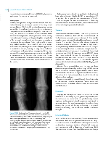
Figure 172.2 Radiographs of the carpus from a 5‐month‐old
Pomeranian with hereditary vitamin D‐resistant rickets. There is Inherited Rickets
substantial widening of the radial and ulnar physes, irregularity of
the physeal–metaphyseal interface and cupping of the Inherited forms of rickets resulting from inborn errors in
metaphyses. The metaphyseal‐epiphyseal bone of the radius and metabolism have been diagnosed in both cats and dogs.
ulna is flared. Source: Reproduced with permission from LeVine There is confusion in the literature with regard to articles
DN, Zhou Y, Ghiloni R, et al. Hereditary 1,25 dihydroxyvitamin on inherited rickets; a number of reported cases are
D‐resistant rickets in a Pomeranian dog caused by a novel
mutation in the vitamin D receptor gene. J Vet Intern Med 2009; more consistent with a diagnosis of nutritional FOD
23: 1278–83. than inherited rickets. Care should be taken in making a