
Page 1594 - Clinical Small Animal Internal Medicine
P. 1594
1532 Section 13 Diseases of Bone and Joint
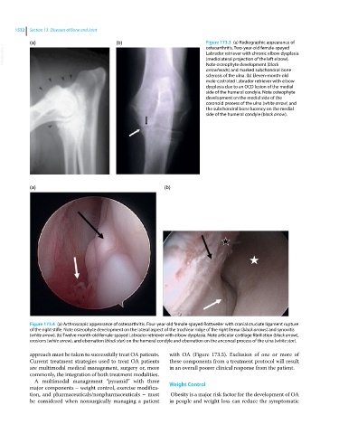
(a) (b) Figure 173.3 (a) Radiographic appearance of
VetBooks.ir Labrador retriever with chronic elbow dysplasia
osteoarthritis. Two‐year‐old female‐spayed
(mediolateral projection of the left elbow).
Note osteophyte development (black
arrowheads) and marked subchondral bone
sclerosis of the ulna. (b) Eleven‐month‐old
male‐castrated Labrador retriever with elbow
dysplasia due to an OCD lesion of the medial
side of the humeral condyle. Note osteophyte
development on the medial side of the
coronoid process of the ulna (white arrow) and
the subchondral bone lucency on the medial
side of the humeral condyle (black arrow).
(a) (b)
Figure 173.4 (a) Arthroscopic appearance of osteoarthritis. Four‐year‐old female‐spayed Rottweiler with cranial cruciate ligament rupture
of the right stifle. Note osteophyte development on the lateral aspect of the trochlear ridge of the right femur (black arrows) and synovitis
(white arrow). (b) Twelve‐month‐old female‐spayed Labrador retriever with elbow dysplasia. Note articular cartilage fibrillation (black arrow),
erosions (white arrow), and ebernation (black star) on the humeral condyle and ebernation on the anconeal process of the ulna (white star).
approach must be taken to successfully treat OA patients. with OA (Figure 173.5). Exclusion of one or more of
Current treatment strategies used to treat OA patients these components from a treatment protocol will result
are multimodal medical management, surgery or, more in an overall poorer clinical response from the patient.
commonly, the integration of both treatment modalities.
A multimodal management “pyramid” with three
major components – weight control, exercise modifica- Weight Control
tion, and pharmaceuticals/nonpharmaceuticals – must Obesity is a major risk factor for the development of OA
be considered when nonsurgically managing a patient in people and weight loss can reduce the symptomatic