
Page 1603 - Clinical Small Animal Internal Medicine
P. 1603
174 Developmental Orthopedic Diseases 1541
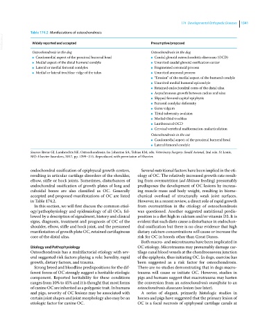
Table 174.2 Manifestations of osteochondrosis
VetBooks.ir Widely reported and accepted Presumptive/proposed
Osteochondrosis in the dog Osteochondrosis in the dog
● Caudomedial aspect of the proximal humeral head ● Caudal glenoid osteochondritis diseccans (OCD)
● Medial aspect of the distal humeral condyle ● Ununited caudal glenoid ossification center
● Lateral or medial femoral condyles ● Fragmented coronoid process
● Medial or lateral trochlear ridge of the talus ● Ununited anconeal process
● “Erosion” of the medial aspect of the humeral condyle
● Ununited medial humeral epicondyle
● Retained endochondral cores of the distal ulna
● Asynchronous growth between radius and ulna
● Slipped femoral capital epiphysis
● Femoral condylar deformity
● Genu valgum
● Tibial tuberosity avulsion
● Medial tibial trochlea
● Lumbosacral OCD
● Cervical vertebral malformation‐malarticulation
Osteochondrosis in the cat
● Caudomedial aspect of the proximal humeral head
● Lateral femoral condyle
Source: Breur GJ, Lambrechts NE. Osteochondrosis. In: Johnston SA, Tobias KM, eds. Veterinary Surgery: Small Animal, 2nd edn. St Louis,
MO: Elsevier Saunders, 2017, pp. 1299–315. Reproduced with permission of Elsevier.
endochondral ossification of epiphyseal growth centers, Several nutritional factors have been implied in the eti-
resulting in articular cartilage disorders of the shoulder, ology of OC. The relatively increased growth rate result-
elbow, stifle or hock joints. Sometimes, disturbances of ing from overnutrition (ad libitum feeding) presumably
endochondral ossification of growth plates of long and predisposes the development of OC lesions by increas-
cuboidal bones are also classified as OC. Generally ing muscle mass and body weight, resulting in biome-
accepted and proposed manifestations of OC are listed chanical overload of structurally weak joint surfaces.
in Table 174.2. However, in a recent review, a direct role of rapid growth
In this section, we will first discuss the common etiol- from overnutrition in the etiology of osteochondrosis
ogy/pathophysiology and epidemiology of all OCs, fol- was questioned. Another suggested nutritional predis-
lowed by a description of signalment, history and clinical position is a diet high in calcium and/or vitamin D3. It is
signs, diagnosis, treatment and prognosis of OC of the evident that such diets cause a disturbance in endochon-
shoulder, elbow, stifle and hock joint, and the presumed dral ossification but there is no clear evidence that high
manifestation of growth plate OC, retained cartilaginous dietary calcium concentrations will cause or increase the
core of the distal ulna. risk for OC in breeds other than Great Danes.
Both macro‐ and microtrauma have been implicated in
Etiology and Pathophysiology OC etiology. Microtrauma may presumably damage car-
Osteochondrosis has a multifactorial etiology with sev- tilage canal blood vessels at the chondroosseous junction
eral suggested risk factors playing a role: heredity, rapid of the epiphysis, thus initiating OC. In dogs, exercise has
growth, dietary factors, and trauma. been suggested as a risk factor for osteochondrosis.
Strong breed and bloodline predispositions for the dif- There are no studies demonstrating that in dogs macro-
ferent forms of OC strongly suggest a heritable etiologic trauma will cause or initiate OC. However, studies in
component. Reported heritability for these conditions pigs and humans suggest that macrotrauma may hasten
ranges from 10% to 45% and it is thought that most forms the conversion from an osteochondrosis manifesta to an
of canine OC are inherited as a polygenic trait. In humans osteochondrosis dissecans lesion (see later).
and pigs, severity of OC lesions may be associated with A series of elegant, primarily histologic studies in
certain joint shapes and joint morphology also may be an horses and pigs have suggested that the primary lesion of
etiologic factor for canine OC. OC is a focal necrosis of epiphyseal cartilage canals at