
Page 617 - Clinical Small Animal Internal Medicine
P. 617
54 Exocrine Pancreatic Insufficiency in Dogs and Cats 585
might be seen. Other possible clinical signs include
VetBooks.ir coprophagia, borborygmus, flatulence, abdominal dis-
comfort, and a poor hair coat. In some cases, EPI may be
subclinical and those cases can only be diagnosed with
appropriate laboratory testing (see section on Trypsin‐
Like Immunoreactivity). Dogs may remain in the sub-
clinical phase for years or sometimes for life.
Cats with EPI have a similar presentation to dogs. In a
retrospective study of 150 cases of feline EPI, the most
common clinical sign was weight loss (which was often
the only clinical sign), followed by loose stools, poor hair
coat, anorexia, increased appetite, depression, watery
diarrhea, and vomiting. It is likely that many of these
clinical signs reflect concurrent diseases such as chronic
pancreatitis and inflammatory bowel disease. In cases
where chronic pancreatitis is the cause of EPI, polyuria
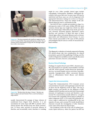
Figure 54.1 This dog presented with significant weight loss due
to EPI. The dog had lost 10 kg during the last few months. This is a and polydipsia may be seen as a result of concurrent dia-
common clinical presentation of dogs with EPI although weight betes mellitus.
loss is often not as severe.
Diagnosis
The diagnostic evaluation of animals suspected of having
EPI should always take into consideration the clinical
presentation, general clinicopathologic findings, and the
results of diagnostic modalities used to specifically assess
pancreatic structure, function, and pathology.
Routine Clinical Pathology
Findings of complete blood count (CBC), chemistry pro-
file, and urinalysis are typically unremarkable in animals
with EPI. When present, routine blood test abnormali-
ties (e.g., increased hepatic enzyme activities, hypoalbu-
minemia, hyperglycemia) reflect concurrent diseases
such as inflammatory bowel disease and diabetes
mellitus.
Trypsin‐like Immunoreactivity
Trypsin‐like immunoreactivity (TLI) measures serum
concentrations of trypsin and trypsinogen and is the test
of choice for the diagnosis of EPI in dogs. This test is
highly sensitive and specific for the diagnosis of EPI.
Animals with EPI produce and secrete lower than normal
Figure 54.2 The feces from the dog in Picture 1. The feces are amounts of trypsinogen in serum and therefore a sub-
large in volume and semi-formed. This is a common finding in
dogs with EPI. normal TLI concentration (usually defined as <2.5 μg/L)
in an animal with compatible clinical signs is sufficient to
make a diagnosis of EPI. A TLI result well within the ref-
usually characterized by passage of large volumes of erence range is sufficient for excluding EPI in the vast
semiformed feces (Figure 54.2). However, it is not majority of cases, and a normal TLI result should direct
uncommon for some dogs with EPI to present with a clinicians towards the investigation of other disorders
clinical picture that deviates from the classic presenta- as the cause of the observed clinical signs.
tion. In those cases, periods of anorexia, absence of One exception to this rule is the case of isolated pan-
diarrhea, and occasionally watery diarrhea or vomiting creatic lipase deficiency, where the patient has EPI but