
Page 731 - Clinical Small Animal Internal Medicine
P. 731
64 Canine Inflammatory Liver Disease 699
biopsy taken at laparoscopy or laparotomy because trucut Therapy
VetBooks.ir biopsies produce misleading results 50% of the time and Therapy for CH is aimed at the underlying cause, if known.
also carry an increased risk of haemorrhage. Needle aspi-
However, as the etiology of most cases is currently not
ration cytology is not reliable for the diagnosis of CH and
is therefore not recommended. understood, specific therapies are usually not possible.
Hepatocytes have a remarkable capacity for regeneration,
The only real absolute contraindication for liver biopsy
is severe coagulopathy. Prior to biopsy, coagulation sta- which means that early diagnosis and therapy have the
potential to reverse disease mechanisms. It is also impor-
tus should be checked, by way of platelet count, meas- tant to initiate therapy as early as possible in an attempt to
urement of PT, PTT, and ideally thromboelastography. inhibit fibrosis, which will ultimately lead to functional
In humans, thromboelastography can predict bleeding impairment. Therapy aimed at addressing clinical signs of
tendencies in cirrhotic patients, but data does not exist liver disease, including ascites, GI ulceration leading to
in dogs to determine how reliable this is as an assessment melena, and hepatic encephalopathy, is also an important
of bleeding tendencies. However, assessment of coagula- part of therapy of the dog with CH. Copper chelators are
tion status should not be a substitute for proper tech- important in dogs with copper-associated hepatitis.
nique and adequate postoperative care. If coagulation Careful dietary management to support the liver is essen-
times are prolonged, vitamin K can be given by injection tial too. Unfortunately, in veterinary medicine there is a
24 hours prior to biopsy and then coagulation times lack of controlled studies on clinical efficacy and pharma-
retested. An alternative approach, if available, is the use cokinetics of the drugs commonly used in canine CH. As
of fresh‐frozen plasma to assist in replenishing deficient a result, many of our current therapeutic protocols are
coagulation factors. All animals should be hospitalised either derived from human hepatology or anecdotal
and monitored carefully for signs of hemorrhage for at reports or originate from low‐quality veterinary clinical
least 12 hours after biopsy. studies. The following refers to the recommended therapy
As detailed previously, standardized criteria for the his-
tologic diagnosis of CH exist. The standard histochemi- of a dog with CH.
cal stain used in the assessment of liver tissue is
hematoxylin and eosin, but consideration should be Corticosteroids
given to the use of additional stains for specific features Corticosteroids have antiinflammatory, immune‐mod-
such as reticulin (connective tissue), Perls’ Prussian blue ulating, and antifibrotic properties. They have a potent
(ferritin), Fouchet’s (bile pigments) and periodic acid– indirect antifibrotic action via reducing prostaglandin
Schiff (polysaccharides). In addition, specific histo- and leukotriene production from inflammatory cells,
chemical stains for copper, such as rubeanic acid or and a weak direct antifibrotic action by inhibiting
rhodanine, should be consider in at‐risk breeds (see ear- mRNA and enzymes. Corticosteroids are indicated in
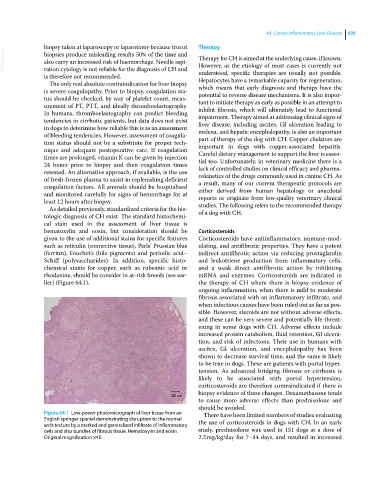
lier) (Figure 64.1). the therapy of CH where there is biopsy evidence of
ongoing inflammation, when there is mild to moderate
fibrosis associated with an inflammatory infiltrate, and
when infectious causes have been ruled out as far as pos-
sible. However, steroids are not without adverse effects,
and these can be very severe and potentially life‐threat-
ening in some dogs with CH. Adverse effects include
increased protein catabolism, fluid retention, GI ulcera-
tion, and risk of infections. Their use in humans with
ascites, GI ulceration, and encephalopathy has been
shown to decrease survival time, and the same is likely
to be true in dogs. These are patients with portal hyper-
tension. As advanced bridging fibrosis or cirrhosis is
likely to be associated with portal hypertension,
corticosteroids are therefore contraindicated if there is
biopsy evidence of these changes. Dexamethasone tends
100 m
to cause more adverse effects than prednisolone and
should be avoided.
Figure 64.1 Low‐power photomicrograph of liver tissue from an There have been limited numbers of studies evaluating
English springer spaniel demonstrating disruption to the normal the use of corticosteroids in dogs with CH. In an early
architecture by a marked and generalized infiltrate of inflammatory
cells and also bundles of fibrous tissue. Hematoxylin and eosin. study, prednisolone was used in 151 dogs at a dose of
Original magnification ×40. 2.2 mg/kg/day for 7–14 days, and resulted in increased