
Page 740 - Clinical Small Animal Internal Medicine
P. 740
708 Section 7 Diseases of the Liver, Gallbladder, and Bile Ducts
ples are labile, and must be placed on ice and analyzed rule out the possibility of liver disease. Many dogs and
VetBooks.ir within 30 minutes to ensure accurate results. Hepatic possibly cats without sonographic abnormalities may still
have important liver abnormalities on histopathology.
function tests are commonly altered in dogs with cir
rhosis, but as cats uncommonly develop significant
cirrhosis. In a study evaluating ultrasound findings with
fibrosis, function can remain normal in this species. Ultrasound is also not a reliable method to diagnose
For further details of laboratory testing in patients histopathologic diagnoses, there were no sonographic
with liver disease, the reader is referred to Chapter 60. features significantly associated with cirrhosis, and it was
concluded that ultrasound could not reliably be used to
detect the presence, absence, or severity of fibrosis. In
Imaging
fact some animals with cirrhosis had ultrasonographi
Imaging modalities most commonly utilized for further cally normal appearing livers. But sonographic findings
evaluation of cirrhosis and its complications include may still be useful in suggesting the possibility of cirrhosis
abdominal radiographs and abdominal ultrasound. and its consequences. Findings that would be most sug
Abdominal radiographs are the most commonly avail gestive of cirrhosis and subsequent PH include identifica
able imaging modality, but are relatively nonspecific for tion of multiple aPSS, a small liver with irregular margins,
hepatic disease. Cirrhosis may result in microhepatica, presence of ascites, and a hyperechoic echotexture. But
which can be visualized on abdominal radiographs. ultimately, biopsy and histopathology are required to
However, this is a change that may be seen in other obtain a definitive diagnosis of cirrhosis.
hepatic diseases such as congenital PSS. Ascites sec Other imaging modalities may also be useful in diag
ondary to PH or hypoalbuminemia may be visible radi nosing the cause or complications of cirrhosis. Computed
ographically as loss of abdominal serosal detail. tomography (CT) and magnetic resonance imaging
Abdominal ultrasound is a very useful noninvasive (MRI) may provide a more complete evaluation of hepatic
imaging modality that can be utilized to further diagnose vasculature than abdominal ultrasound. When utilized
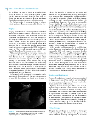
liver disease and its complications (Figure 65.2). with contrast, CT and MRI may also provide further eval
Ultrasound can evaluate hepatic parenchymal echo uation regarding location of neoplasia and possible
genicity and uniformity, overall hepatic size, biliary metastasis. Rectal or transsplenic nuclear scintigraphy
structures, hepatic and portal vessels, and identify very can be useful in the diagnosis of PSS, when ultrasound is
small volumes of ascites that might not be visible radio inconclusive, although scintigraphy may not be able to
graphically. It can also be utilized to identify multiple distinguish congenital versus aPSS. Further details of
aPSS . Ultrasound is also useful in guiding safe collection diagnostic imaging in the investigation of cirrhosis and its
of samples of ascites or bile for fluid analysis, or ultra complications can be found in Chapter 61.
sound‐guided “Tru‐Cut” biopsies.
Unfortunately, while ultrasound is a very useful nonin
vasive test, it does not identify changes pathognomonic Cytology and Fluid Analysis
for a specific liver disease, and should never be used to Fine needle aspiration cytology is an inadequate method
to diagnose cirrhosis. While cytology may help identify
the underlying cause of cirrhosis, it cannot confirm the
presence or absence of fibrous tissue
Cytology of ascitic fluid may be utilized when attempt
ing to identify whether the ascites is caused by PH, infec
tious disease, or neoplastic disease. Typically, ascites
secondary to PH is a modified transudate (also refereed to
as a proteinrich transudate), or possibly a pure transu
date. PH ascites is nonneoplastic, and noninflammatory
in origin. Cytology and culture of bile may also be impor
tant in diagnosing the underlying cause of cirrhosis. Biliary
cultures have been shown to yield a significantly higher
rate of positive growth than hepatic tissue cultures.
Histopathology
The gold standard diagnosis of cirrhosis is liver biopsy
Figure 65.2 Ultrasonographic image of a cirrhotic liver. Note the
presence of large‐volume ascites, a small liver with irregular with histopathology that identifies the presence and
hepatic margination, and mottled echogenicity. extent of fibrosis. Not only is it important to document