Page 810 - Clinical Small Animal Internal Medicine
P. 810
778  Section 8  Neurologic Disease

               excess salt intake: salt poisoning, administration of IV   with normovolemic or hypervolemic hyponatremia
            ●
  VetBooks.ir  hypertonic solutions (NaCl), sodium bicarbonate, or   associated with excessive water intake or renal reten-
              saline emetics
                                                              tion, water restriction (i.e., limiting water intake to less
               insufficient water intake: lack of access, inability to
            ●
              drink, CNS disease resulting in primary adipsia (absence   than urine output) can be performed. In instances where
                                                              severe neurologic signs are seen associated with normal
              of thirst), mental depression, congenital adipsia.  or excessive intravascular fluid, frusemide (2–4 mg/kg
                                                              IV) can be used to promote renal water excretion.
            Marked hypernatremia may induce cerebral signs,     Chronic hyponatremia may be more difficult to treat
            such as depression, weakness, irritability, uncharacteris-  since overrapid correction of the deficit can result in a
            tic aggression, confusion, propulsive circling, demen-  worsening of clinical signs associated with central pon-
            tia,  seizures, coma, and death as the result of cellular   tine myelinolysis. Lesions appear in white matter
            dehydration of neurons. Neurologic signs may not   associated with death of oligodendrocytes and loss of
            occur  until serum Na levels exceed 170–175 mEq/L   myelin, resulting in bilaterally symmetric lesions in the
            (>350 mOsm/kg), although the severity of neurologic   pons and rarely (10% of cases) thalamus, subthalamic
            signs depends especially on the rate of increase, with   nuclei, striatum, internal capsule, amygdala, lateral
            signs being most severe in animals with rapidly devel-  geniculate body, white matter of the cerebellum and deep
            oping hyperosmolality.                            layers of the cerebral cortex. The postulated hypothesis
              Treatment depends on the rate at which hypernatremia
            develops; overrapid correction can lead to worsening   is  that with chronicity,  cells within  the brain  extrude
                                                              intracellular electrolytes followed by organic osmolytes
            clinical signs (due to a relative hyponatremia and move-  (“idiogenic osmoles”), in order to reduce the risk of brain
            ment of water from the vascular spaces into the brain).   edema. When sodium is replaced intravascularly as part
            Replacement of the water deficit should be undertaken   of fluid therapy, osmotic attraction of water out of cells
            using 5% dextrose with the aim of correction of the deficit   in the brain results in dehydration of brain tissue. Clinical
            over 24 hours (or longer). In animals with acute‐onset   signs occur several days after fluid treatment: ataxia,
            hypernatremia, correction of Na levels at a rate of 1.0   weakness, hypermetria, visual deficits, depressed men-
            mEq/h is acceptable. However, if the hypernatremia is   ace  response (with normal  pupillary light  response
            more chronic, Na levels should not be allowed to change   [PLR]), reduced proprioception, exaggerated licking
            at more than 0.5 mEq/h. In animals with excess salt   movements, episodic myoclonus, whole‐body spasms,
            intake, diuretics should be given in conjunction with fluid   obtundation, and tetraparesis. Prognosis for recovery is
            therapy to avoid pulmonary edema.
                                                              reasonable; in one study of two dogs, one died but the
                                                              other recovered over 4–7 weeks. As a result, it is impor-
            Hyponatremia                                      tant to stick to the guideline of replacing Na at a rate no
            Hyponatremia occurs when serum Na levels are less than   greater than 0.5 mEq/L/h.
            146 mEq/L in dogs (151 mEq/L in cats). Hyponatremia
            can be further divided into true hyponatremia (associ-
            ated with total body water in excess of Na – serum osmo-    Neoplastic Causes
            lality is low, <290 mOsm/L) and  pseudohyponatremia
            (low Na with normal or increased serum osmolality).  Brain Tumors
             Clinical signs of hyponatremia relate primarily to the
            rate of onset of hyponatremia. In acute hyponatremia,   Tumors affecting the brain can arise as one of several
            sodium levels within the brain are relatively normal   forms.
            while sodium levels within the vascular spaces are low.     Primary tumors arise from cells and structures of the
            Water flows down its concentration gradient into the   ●  brain itself.
            brain, producing cerebral edema and raised intracranial     Secondary tumors arise from cells and structures adja-
            pressure (ICP). Signs include generalized weakness,   ●  cent to the brain.
            depression, stupor, coma, seizures, and dementia.     Metastatic tumors arise from spread of a tumor at a
            Treatment is aimed at increasing sodium levels: use   ●  distant site.
            hypertonic saline (3–5%) when serum Na <115 mEq/L.
            The Na deficit is calculated using:               The most common tumors that affect dogs (and humans)
                                                              are primary tumors. They occur with a slightly greater inci-
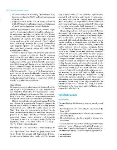
            Defect mEq /l  140  measured Na  bodyweight kg  . 03  dence than in people (14–15 per 100 000 dogs vs 8–9 per

            The replacement fluid should be given slowly over   100 000 people at risk). The most common types include:
            12–24 hours. For animals with hypovolemic hypona-  ●   meningiomas
            tremia, normal isotonic saline can be used. For animals   ●   astrocytomas

