Page 844 - Clinical Small Animal Internal Medicine
P. 844
812  Section 8  Neurologic Disease

                                                              or orthopedic abnormalities secondary to the effects of
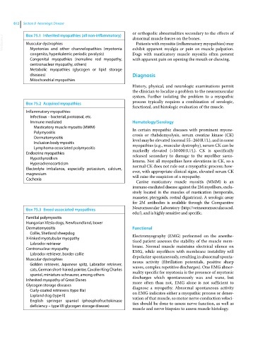
             Box 75.1  Inherited myopathies (all non-inflammatory)
  VetBooks.ir  Muscular dystrophies                           abnormal muscle forces on the bones.
                                                                Patients with myositis (inflammatory myopathies) may
               Myotonias and other channelopathies (myotonia
                                                              Dogs with masticatory  muscle myositis often present
                 congenita, hyperkalemic periodic paralysis)  exhibit apparent myalgia or pain on muscle palpation.
               Congenital myopathies (nemaline rod myopathy,   with apparent pain on opening the mouth or chewing.
               centronuclear myopathy, others)
               Metabolic myopathies (glycogen or lipid storage
               diseases)                                        Diagnosis
               Mitochondrial myopathies
                                                              History, physical, and neurologic examinations permit
                                                              the clinician to localize a problem to the neuromuscular
                                                              system.  Further  isolating  the  problem  to  a  myopathic
             Box 75.2  Acquired myopathies                    process typically requires a combination of serologic,
                                                              functional, and histologic evaluation of the muscle.
             Inflammatory myopathies
               Infectious – bacterial, protozoal, etc.
               Immune mediated                                Hematology/Serology
                 Masticatory muscle myositis (MMM)            In certain myopathic diseases with prominent myone-
                 Polymyositis                                 crosis or rhabdomyolysis, serum creatine  kinase (CK)
                 Dermatomyositis                              level may be elevated (normal 55–260 IU/L), and in some
                 Inclusion body myositis                      myopathies (e.g., muscular dystrophy), serum CK can be
                 Lymphoma‐associated polymyositis
             Endocrine myopathies                             markedly elevated (>50 000 IU/L). CK is specifically
                                                              released secondary to damage to the myofiber sarco-
               Hypothyroidism                                 lemma. Not all myopathies have elevations in CK, so a
               Hyperadrenocorticism
             Electrolyte imbalance, especially potassium, calcium,   normal CK does not rule out a myopathic process; how-
                                                              ever, with appropriate clinical signs, elevated serum CK
             magnesium                                        will raise the suspicion of a myopathy.
             Cachexia
                                                                Canine  masticatory  muscle  myositis  (MMM)  is  an
                                                              immune‐mediated disease against the 2M myofibers, exclu-
                                                              sively located in the muscles of mastication (temporalis,
                                                              masseter, pterygoids, rostral digastricus). A   serologic assay
                                                              for 2M antibodies is available through the Comparative
             Box 75.3  Breed‐associated myopathies            Neuromuscular Laboratory (http://vetneuromuscular.ucsd.
                                                              edu/), and is highly sensitive and specific.
             Familial polymyositis
             Hungarian Vizsla dogs, Newfoundland, boxer
             Dermatomyositis                                  Functional
               Collie, Shetland sheepdog
             X‐linked myotubular myopathy                     Electromyography (EMG) performed on the anesthe-
               Labrador retriever                             tized patient assesses the stability of the muscle mem-
             Centronuclear myopathy                           brane. Normal muscle maintains electrical silence on
               Labrador retriever, border collie              EMG, while myofibers with membrane instability will
             Muscular dystrophies                             depolarize spontaneously, resulting in abnormal sponta-
               Golden retriever, Japanese spitz, Labrador retriever,   neous  activity  (fibrillation  potentials,  positive  sharp
               cats, German short‐haired pointer, Cavalier King Charles   waves, complex repetitive discharges). One EMG abnor-
               spaniel, miniature schnauzer, among others     mality specific for myotonia is the presence of myotonic
             Inherited myopathy of Great Danes                discharges which spontaneously wax and wane, but
             Glycogen storage diseases                        more  often  than  not,  EMG  alone  is  not  sufficient  to
               Curly‐coated retrievers (type IIIa)            diagnose  a  myopathy.  Abnormal  spontaneous  activity
               Lapland dog (type II)                          on EMG indicates either a myopathic process or dener-
               English springer  spaniel (phosphofructokinase   vation of that muscle, so motor nerve conduction veloci-
                 deficiency – type VII glycogen storage disease)  ties should be done to assess nerve function, as well as
                                                              muscle and nerve biopsies to assess muscle histology.

