
Page 979 - Clinical Small Animal Internal Medicine
P. 979
917
VetBooks.ir
95
Wolbachia pipientis Infection
Pedro P. Vissotto de Paiva Diniz, DVM, PhD
College of Veterinary Medicine, Western University of Health Sciences, Pomona, CA, USA
Etiology/Pathophysiology in inflammatory lesions and pulmonary artery vasocon-
striction. Experimental infection with D. immitis free of
Wolbachia pipientis is a gram‐negative bacteria belonging Wolbachia results in a reduced inflammatory response
to the family Anaplasmataceae. The organism is found in after anthelminthic therapy. Furthermore, antibiotic
all developmental stages of Dirofilaria immitis, the filarial therapy that targets Wolbachia results in a significant
helminth that causes heartworm disease in dogs and cats. reduction in inflammatory reactions when anthelmintic
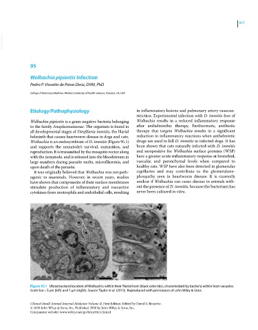
Wolbachia is an endosymbiont of D. immitis (Figure 95.1) drugs are used to kill D. immitis in infected dogs. It has
and supports the nematode’s survival, maturation, and been shown that cats naturally infected with D. immitis
reproduction. It is transmitted by the mosquito vector along and seropositive for Wolbachia surface proteins (WSP)
with the nematode, and is released into the bloodstream in have a greater acute inflammatory response at bronchial,
large numbers during parasite molts, microfilaremia, and vascular, and parenchymal levels when compared to
upon death of the parasite. healthy cats. WSP have also been detected in glomerular
It was originally believed that Wolbachia was not path- capillaries and may contribute to the glomerulone-
ogenic to mammals. However, in recent years, studies phropathy seen in heartworm disease. It is currently
have shown that components of their surface membranes unclear if Wolbachia can cause disease in animals with-
stimulate production of inflammatory and vasoactive out the presence of D. immitis, because the bacterium has
cytokines from neutrophils and endothelial cells, resulting never been cultured in vitro.
Figure 95.1 Ultrastructural location of Wolbachia within their filarial host (black asterisks), characterized by bacteria within host vacuoles.
Scale bar = 5 μm (left) and 1 μm (right). Source: Taylor et al. (2013). Reproduced with permission of John Wiley & Sons.
Clinical Small Animal Internal Medicine Volume II, First Edition. Edited by David S. Bruyette.
© 2020 John Wiley & Sons, Inc. Published 2020 by John Wiley & Sons, Inc.
Companion website: www.wiley.com/go/bruyette/clinical