
Page 358 - The Toxicology of Fishes
P. 358
338 The Toxicology of Fishes
liver from trout (Oncorhynchus mykiss) and channel catfish (Ictalurus punctatus) have revealed numerous
transitional passageways between the canaliculi of hepatocytes and biliary ductules (Hampton et al.,
1988). Transitional passageways (Figure 7.8A), the bile preductules (Hinton and Pool, 1976), are sites
where hepatocytes and biliary epithelial cells with oval perikarya share junctional complexes (Hampton
et al., 1988). This terminology in fish was adopted based on the flow of bile and the finding of transitional
cells in rodent liver between epithelial cells of cholangioles and hepatocytes (Steiner and Carruthers,
1961). Biliary epithelial cells between canaliculi and ductules or cholangioles (completely lined by
biliary epithelial cells) were designated in fish as bile preductular epithelial cells (BPDECs) (Figure
7.8). These small, oval cells form junctional complexes with both hepatocytes and adjacent bile ductular
epithelial cells. The fine structure of these cells includes scant cytoplasm, intermediate filaments, and
an absence of a basal lamina (Hampton et al., 1988). Hampton et al. (1988) extended their observations
to bile ductules and ducts in control rainbow trout. As the diameters of bile preductules enlarge, additional
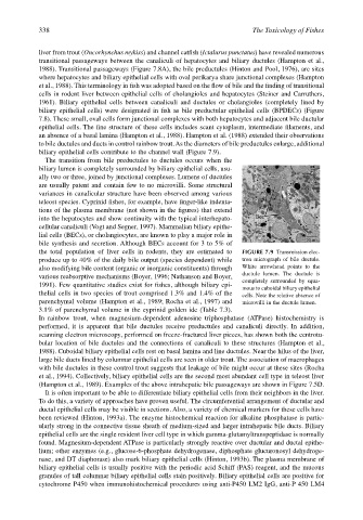
biliary epithelial cells contribute to the channel wall (Figure 7.9).
The transition from bile preductules to ductules occurs when the
biliary lumen is completely surrounded by biliary epithelial cells, usu-
ally two or three, joined by junctional complexes. Lumens of ductules
are usually patent and contain few to no microvilli. Some structural
variances in canalicular structure have been observed among various
teleost species. Cyprinid fishes, for example, have finger-like indenta-
tions of the plasma membrane (not shown in the figures) that extend
into the hepatocytes and show continuity with the typical interhepato-
cellular canaliculi (Vogt and Segner, 1997). Mammalian biliary epithe-
lial cells (BECs), or cholangiocytes, are known to play a major role in
bile synthesis and secretion. Although BECs account for 3 to 5% of
the total population of liver cells in rodents, they are estimated to FIGURE 7.9 Transmission elec-
produce up to 40% of the daily bile output (species dependent) while tron micrograph of bile ductule.
also modifying bile content (organic or inorganic constituents) through White arrowhead points to the
ductule lumen. The ductule is
various reabsorptive mechanisms (Boyer, 1996; Nathanson and Boyer,
completely surrounded by squa-
1991). Few quantitative studies exist for fishes, although biliary epi-
mous to cuboidal biliary epithelial
thelial cells in two species of trout comprised 1.3% and 1.4% of the cells. Note the relative absence of
parenchymal volume (Hampton et al., 1989; Rocha et al., 1997) and microvilli in the ductule lumen.
3.1% of parenchymal volume in the cyprinid golden ide (Table 7.3).
In rainbow trout, when magnesium-dependent adenosine triphosphatase (ATPase) histochemistry is
performed, it is apparent that bile ductules receive preductules and canaliculi directly. In addition,
scanning electron microscopy, performed on freeze-fractured liver pieces, has shown both the centrotu-
bular location of bile ductules and the connections of canaliculi to these structures (Hampton et al.,
1988). Cuboidal biliary epithelial cells rest on basal lamina and line ductules. Near the hilus of the liver,
large bile ducts lined by columnar epithelial cells are seen in older trout. The association of macrophages
with bile ductules in these control trout suggests that leakage of bile might occur at these sites (Rocha
et al., 1994). Collectively, biliary epithelial cells are the second most abundant cell type in teleost liver
(Hampton et al., 1989). Examples of the above intrahepatic bile passageways are shown in Figure 7.5D.
It is often important to be able to differentiate biliary epithelial cells from their neighbors in the liver.
To do this, a variety of approaches have proven useful. The circumferential arrangement of ductular and
ductal epithelial cells may be visible in sections. Also, a variety of chemical markers for these cells have
been reviewed (Hinton, 1993a). The enzyme histochemical reaction for alkaline phosphatase is partic-
ularly strong in the connective tissue sheath of medium-sized and larger intrahepatic bile ducts. Biliary
epithelial cells are the single resident liver cell type in which gamma-glutamyltranspeptidase is normally
found. Magnesium-dependent ATPase is particularly strongly reactive over ductular and ductal epithe-
lium; other enzymes (e.g., glucose-6-phosphate dehydrogenase, diphosphate glucuronosyl dehydroge-
nase, and DT diaphorase) also mark biliary epithelial cells (Hinton, 1993b). The plasma membrane of
biliary epithelial cells is usually positive with the periodic acid Schiff (PAS) reagent, and the mucous
granules of tall columnar biliary epithelial cells stain positively. Biliary epithelial cells are positive for
cytochrome P450 when immunohistochemical procedures using anti-P450 LM2 IgG, anti-P 450 LM4