Page 216 - Veterinary Histology of Domestic Mammals and Birds, 5th Edition
P. 216
198  Veterinary Histology of Domestic Mammals and Birds


       VetBooks.ir


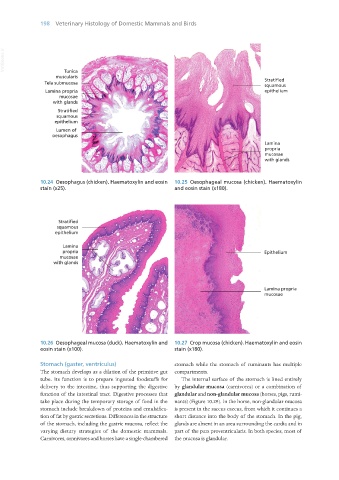
                                                                                                  Stratified
                                                                                                  squamous
                                                                                                  epithelium


                                                                                                  Lamina
                                                                                                  propria
                                                                                                  mucosae
                                                                                                  with glands


                   10.24  Oesophagus (chicken). Haematoxylin and eosin   10.25  Oesophageal mucosa (chicken). Haematoxylin
                   stain (x25).                                   and eosin stain (x180).


                   10.26  Oesophageal mucosa (duck). Haematoxylin and   10.27  Crop mucosa (chicken). Haematoxylin and eosin
                   eosin stain (x100).                            stain (x180).


                   Stomach (gaster, ventriculus)                  stomach while the stomach of ruminants has multiple
                   The stomach develops as a dilation of the primitive gut  compartments.
                   tube. Its function is to prepare ingested foodstuffs for   The internal surface of the stomach is lined entirely
                   delivery to the intestine, thus supporting the digestive  by glandular mucosa (carnivores) or a combination of
                   function of the intestinal tract. Digestive processes that  glandular and non-glandular mucosa (horses, pigs, rumi-
                   take place during the temporary storage of food in the  nants) (Figure 10.29). In the horse, non-glandular mucosa
                   stomach include breakdown of proteins and emulsifica-  is present in the saccus caecus, from which it continues a
                   tion of fat by gastric secretions. Differences in the structure  short distance into the body of the stomach. In the pig,
                   of the stomach, including the gastric mucosa, reflect the  glands are absent in an area surrounding the cardia and in
                   varying dietary strategies of the domestic mammals.  part of the pars proventricularis. In both species, most of
                   Carnivores, omnivores and horses have a single-chambered  the mucosa is glandular.


       Vet Histology.indb   198                                                                                  16/07/2019   15:00

