
Page 286 - Veterinary Histology of Domestic Mammals and Birds, 5th Edition
P. 286
268 Veterinary Histology of Domestic Mammals and Birds
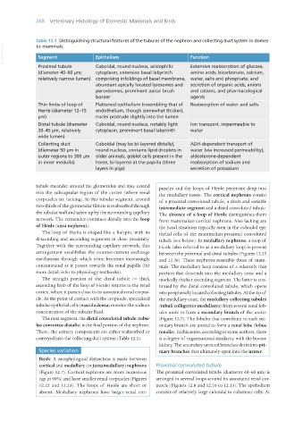
Table 12.1 Distinguishing structural features of the tubules of the nephron and collecting duct system in domes-
VetBooks.ir Segment Epithelium Function
tic mammals.
Proximal tubule Cuboidal, round nucleus, acidophilic Extensive reabsorption of glucose,
(diameter 40–60 μm; cytoplasm, extensive basal labyrinth amino acids, bicarbonate, calcium,
relatively narrow lumen) comprising infoldings of basal membrane, water, salts and phosphate, and
abundant apically located lysosomes and secretion of organic acids, anions
peroxisomes, prominent apical brush and cations, and pharmacological
border agents
Thin limbs of loop of Flattened epithelium (resembling that of Reabsorption of water and salts
Henle (diameter 12–15 endothelium, though somewhat thicker),
μm) nuclei protrude slightly into the lumen
Distal tubule (diameter Cuboidal, round nucleus, notably light Ion transport, impermeable to
30–45 μm, relatively cytoplasm, prominent basal labyrinth water
wide lumen)
Collecting duct Cuboidal (may be bi-layered distally), ADH-dependent transport of
(diameter 50 μm in round nucleus, contains lipid droplets in water (via increased permeability),
outer regions to 300 μm older animals, goblet cells present in the aldosterone-dependent
in inner medulla) horse, bi-layered at the papilla (three reabsorption of sodium and
layers in pigs) secretion of potassium
tubule meander around the glomerulus and may extend puscles and the loops of Henle penetrate deep into
into the subcapsular region of the cortex (where renal the medullary tissue. The cortical nephrons consist
corpuscles are lacking). In this tubular segment, around of a proximal convoluted tubule, a short and variable
two-thirds of the glomerular filtrate is reabsorbed through intermediate segment and a distal convoluted tubule.
the tubular wall and taken up by the surrounding capillary The absence of a loop of Henle distinguishes them
network. The remainder continues distally into the loop from mammalian cortical nephrons. Also lacking are
of Henle (ansa nephroni). the basal striations typically seen in the cuboidal epi-
The loop of Henle is shaped like a hairpin, with its thelial cells of the mammalian proximal convoluted
descending and ascending segments in close proximity. tubule (see below). In medullary nephrons, a loop of
Together with the surrounding capillary network, this Henle (also referred to as a medullary loop) is present
arrangement establishes the counter-current exchange between the proximal and distal tubules (Figures 12.25
mechanism through which urine becomes increasingly and 12.26). These nephrons resemble those of mam-
concentrated as it passes towards the renal papilla (for mals. The medullary loop consists of a relatively thin
more detail, refer to physiology textbooks). portion that descends into the medullary cone and a
The straight portion of the distal tubule (= thick markedly thicker ascending segment. The latter is con-
ascending limb of the loop of Henle) returns to the renal tinued by the distal convoluted tubule, which opens
cortex, where it passes close to its associated renal corpus- into peripherally located collecting tubules. At the tip of
cle. At the point of contact with the corpuscle, specialised the medullary cone, the medullary collecting tubules
tubular epithelial cells (macula densa) monitor the sodium (tubuli colligentes medullares) from several renal lob-
concentration of the tubular fluid. ules unite to form a secondary branch of the ureter
The next segment, the distal convoluted tubule (tubu- (Figure 12.7). The lobules that contribute to each sec-
lus contortus distalis) is the final portion of the nephron. ondary branch are united to form a renal lobe (lobus
There, the urinary components are either reabsorbed or renalis). In this sense, according to some authors, there
conveyed into the collecting duct system (Table 12.1). is a degree of organisational similarity with the bovine
kidney. The secondary ureteral branches drain into pri-
Species variation mary branches that ultimately open into the ureter.
Birds: A morphological distinction is made between
cortical and medullary (or juxtamedullary) nephrons Proximal convoluted tubule
(Figure 12.7). Cortical nephrons are more numerous The proximal convoluted tubule (diameter 40–60 μm) is
(up to 90%) and have smaller renal corpuscles (Figures arranged in several loops around its associated renal cor-
12.22 and 12.23). The loops of Henle are short or puscle (Figures 12.8 and 12.10 to 12.13). The epithelium
absent. Medullary nephrons have larger renal cor- consists of relatively large cuboidal to columnar cells. At
Vet Histology.indb 268 16/07/2019 15:03