
Page 340 - Veterinary Histology of Domestic Mammals and Birds, 5th Edition
P. 340
322 Veterinary Histology of Domestic Mammals and Birds
shallow primary and secondary folds. Initially the infundib-
− stratum longitudinale,
VetBooks.ir • tunica serosa: ulum is non-glandular (Figure 14.27). Towards the caudal
portion of the funnel, alveolar invaginations (fossae glan-
− epithelium serosae and
dulares infundibuli) appear in the lamina propria.
− lamina propria serosae.
In the subsequent tubular segment, these infundibular
glands increase in size and complexity forming glandulae
The epithelium mucosae is initially simple and flat, then tubi infundibulares. These produce a protein-rich secre-
transitions through cuboidal to pseudostratified columnar. tion that surrounds the oocyte.
The columnar cells consist of intra-epithelial gland cells, The wall of the funnel contains smooth muscle, giving
ciliated surface cells and basal cells. The distal segments it contractile properties that aid in the uptake of oocytes
contain regions of pseudostratified columnar epithelium after ovulation. In the tubular section, the wall of the
that subsequently become reduced in thickness. infundibulum is thicker and features more prominent pri-
The lamina propria mucosae contains glands along mary and secondary folds. Fertilisation of the oocyte by
most of its length. These vary considerably in structure, spermatozoa occurs in this tubular portion.
number and density in different segments of the oviduct. Transit of the egg through the infundibulum takes
Mucosal folds are present to a greater or lesser degree around 15–20 minutes. During its passage, the egg
throughout the oviduct. The height and thickness of the becomes surrounded by glycoproteins secreted by the
folds vary from one segment of the oviduct to another glands. These form the inner dense layer of albumen
(Figures 14.27 to 14.29). The arrangement of the folds in which later gives rise to the twisted chalazae that suspend
a gentle spiral causes the egg to turn slowly around its the yolk as it rotates about its longitudinal axis.
longitudinal axis as it travels through the oviduct.
The circular and longitudinal muscle layers of the Magnum
tunica muscularis bring about peristaltic contractions that The magnum is the longest segment of the oviduct. In the
assist in transporting the egg, and antiperistaltic contrac- chicken, it reaches 34 cm. The magnum follows a loop-
tions that facilitate swift passage of spermatozoa. ing course, resembling that of the jejunum. Within this
segment, the epithelium transitions from pseudostrati-
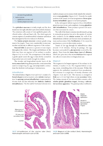
Infundibulum fied columnar to a single layer of mostly columnar cells
The infundibulum (Figures 14.26 and 14.27) consists of a (Figures 14.26 and 14.28). The mucosa is arranged in
funnel-shaped proximal section and a tubular distal por- folds up to 22 mm high (there are no secondary folds).
tion. Its opening (ostium infundibulare) is approximately The lamina propria of the mucosal folds contains coiled
80 mm wide and has relatively few fimbriae (fimbriae tubular glands (glandulae magni), forming a substan-
infundibulares). The thin wall of the funnel is thrown into tial secretory apparatus. The glands produce ovalbumin,
14.27 Infundibulum of the uterine tube (hen). 14.28 Magnum of the oviduct (hen). Haematoxylin
Haematoxylin and eosin stain (x120). and eosin stain (x300).
Vet Histology.indb 322 16/07/2019 15:05