
Page 104 - Veterinary Laser Therapy in Small Animal Practice
P. 104
90 Veterinary Laser Therapy in Small Animal Practice
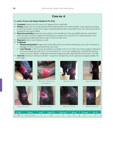
Case no. 6
L., canine, 15 years old, Belgian Shepherd, FS, 23 kg
• Complaint: wound over left carpus and lameness from same limb.
• History: surgery for soft tissue sarcoma at the referring practice 4 months before. Clean margins according
to pathology report. A week after surgery, patient presented with wound dehiscence. The wound size had not
changed for the past 11 weeks.
• Physical examination: lameness was graded as 2/5 and related to the wound after physical examination
2
ruled out other locations. Despite lack of healing, the wound, which was 35 cm in size, presented a well-
vascularized granulation bed with no signs of infection (Fig. C6.1).
• Diagnosis: chronic (non-healing) wound.
• Treatment:
• Wound management: lavaged with sterile saline and covered with sterile gauze and a non-compressive
bandage. No oral or topical treatments were used.
• Laser therapy: on the first day, the patient was treated with 4 J/cm ; 18 h later, the granulation bed had an
2
exuberant appearance (Fig. C6.2), so a lower dose of 2 J/cm was used that day, using half the previous
2
power and power density. Treatments were then performed q48h, and after the second week, q72–96h.
• Outcome: full closure of the wound after 11 sessions in 32 days (Fig. C6.8). Lameness disappeared after the
first week.
Figure C6.1 Initial aspect. Figure C6.2 18 h after first Figure C6.3 Day 7. Figure C6.4 Day 10.
laser treatment.
Figure C6.5 Day 17. Figure C6.6 Day 21. Figure C6.7 Day 28. Figure C6.8 Day 32.
P (W) Tx time J/cm 2 Total J/Tx Spot (cm ) W/cm 2 Tx/week No. Tx
2
a
3 132 s (2.2 min) 4 400 6 0.5 3-2 11
REDONDO PRINT (4-COL BLEED).indd 90 08/08/2019 09:47