
Page 139 - Veterinary Laser Therapy in Small Animal Practice
P. 139
Pointing light at musculoskeletal and neurological conditions: clinical applications 125
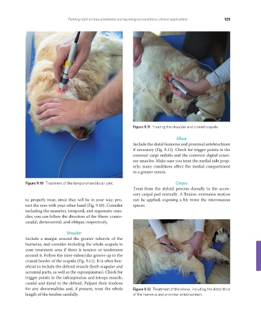
Figure 9.11 Treating the shoulder and cranial scapula.
Elbow
Include the distal humerus and proximal antebrachium
if necessary (Fig. 9.12). Check for trigger points in the
extensor carpi radialis and the common digital exten-
sor muscles. Make sure you treat the medial side prop-
erly; many conditions affect the medial compartment
to a greater extent.
Figure 9.10 Treatment of the temporomandibular joint. Carpus
Treat from the styloid process dorsally to the acces-
sory carpal pad ventrally. A flexion–extension motion
to properly treat, since they will be in your way; pro- can be applied, exposing a bit more the interosseous
tect the eyes with your other hand (Fig. 9.10). Consider spaces.
including the masseter, temporal, and zygomatic mus-
cles; you can follow the direction of the fibers: cranio-
caudal, dorsoventral, and oblique, respectively.
Shoulder
Include a margin around the greater tubercle of the
humerus, and consider including the whole scapula in
your treatment area if there is tension or tenderness
around it. Follow the inter-tubercular groove up to the
cranial border of the scapula (Fig. 9.11). It is often ben-
eficial to include the deltoid muscle (both scapular and
acromial parts, as well as the supraspinatus). Check for
trigger points in the infraspinatus and triceps muscle,
caudal and distal to the deltoid. Palpate their tendons
for any abnormalities and, if present, treat the whole Figure 9.12 Treatment of the elbow, including the distal third
length of the tendon carefully. of the humerus and proximal antebrachium.
REDONDO PRINT (4-COL BLEED).indd 125 08/08/2019 09:48