
Page 46 - Veterinary Laser Therapy in Small Animal Practice
P. 46
32 Veterinary Laser Therapy in Small Animal Practice
experimental burn wounds. [60, 119] Some have com-
pared LT with other modalities: an in vitro experiment
with fibroblasts found laser to be more stimulating
than ultrasound in terms of fibroblast activity [136] and
as good as electrical stimulation in experimental rat
skin incisions – both electrical stimulation and LT
decreased the duration of the inflammatory phase and
increased the number of fibroblasts and the concen-
tration of hydroxyproline compared with their control
groups.
Steroid treatment may affect the efficacy of LT.
One study concluded that LT was useless on open
skin wounds in corticosteroid-treated diabetic rats
(although it did accelerate epithelialization in the
no-steroid group). [137] But using similar parameters,
another research group [118] concluded that LT may
help counteract the inhibitory effect of anti-inflamma-
tory drugs on wound healing: while the groups treated
with a non-steroidal anti-inflammatory drug (NSAID;
celecoxib 22 mg/kg) or corticosteroid (dexamethasone
5 mg/kg) tended to have lower wound cellularity and
more immature granulation, treating those animals
with LT helped restore cellularity to the baseline level,
increasing fibroblast migration, collagen synthesis, and
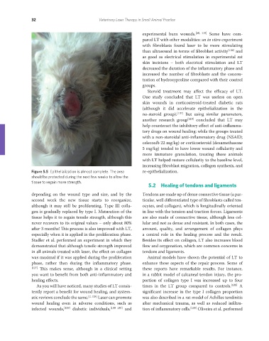
Figure 5.5 Epithelialization is almost complete. The area re-epithelialization.
should be protected during the next few weeks to allow the
tissue to regain more strength.
5.2 Healing of tendons and ligaments
depending on the wound type and size, and by the Tendons are made up of dense connective tissue (a par-
second week the new tissue starts to reorganize, ticular, well differentiated type of fibroblasts called ten-
although it may still be proliferating. Type III colla- ocytes, and collagen), which is longitudinally oriented
gen is gradually replaced by type I. Maturation of the in line with the tension and traction forces. Ligaments
tissue helps it to regain tensile strength, although this are also made of connective tissue, although less cel-
never recovers to its original values – only about 80% lular and not as dense and resistant. In both cases, the
after 3 months! This process is also improved with LT, amount, quality, and arrangement of collagen plays
especially when it is applied in the proliferation phase. a central role in the healing process and the result.
Stadler et al. performed an experiment in which they Besides its effect on collagen, LT also increases blood
demonstrated that although tensile strength improved flow and oxygenation, which are common concerns in
in all animals treated with laser, the effect on collagen tendons and ligaments.
was maximal if it was applied during the proliferation Animal models have shown the potential of LT to
phase, rather than during the inflammatory phase. enhance these aspects of the repair process. Some of
[127] This makes sense, although in a clinical setting these reports have remarkable results. For instance,
you want to benefit from both anti-inflammatory and in a rabbit model of calcaneal tendon injury, the pro-
healing effects. portion of collagen type I was increased up to four
As you will have noticed, many studies of LT consis- times in the LT group compared to controls. [138] A
tently report a benefit for wound healing, and system- significant increase in the type I collagen proportion
atic reviews conclude the same. [2, 128] Laser can promote was also described in a rat model of Achilles tendinitis
wound healing even in adverse conditions, such as after mechanical trauma, as well as reduced infiltra-
infected wounds, [129] diabetic individuals, [130–135] and tion of inflammatory cells. [139] Oliveira et al. performed
REDONDO PRINT (4-COL BLEED).indd 32 08/08/2019 09:47