
Page 525 - The Veterinary Care of the Horse
P. 525
12
VetBooks.ir THE HORSE’S SPINE AND PELVIS
THE HORSE’S SPINE
Anatomy
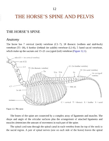
The horse has 7 cervical (neck) vertebrae (C1–7), 18 thoracic (withers and mid-back)
vertebrae (T1–18), 6 lumbar (behind the saddle) vertebrae (L1-6), 5 fused sacral vertebrae,
which make up the sacrum and 15–21 coccygeal (tail) vertebrae (Figure 12.1).
Figure 12.1 The spine
The bones of the spine are connected by a complex array of ligaments and muscles. The
shape and angle of the articular surfaces plus the arrangement of attached ligaments and
muscles determines the amount of movement in each part of the spine.
The spinal cord runs through the spinal canal in each vertebra from the top of the neck to
the sacral region. A pair of spinal nerves (one on each side of the horse) leaves the spinal