
Page 193 - Avian Virology: Current Research and Future Trends
P. 193
186 | Kibenge et al.
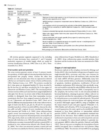
Table 6.1 Continued
Genome Encoded viral protein,
segment and location, and number Calculated
size (bp) of units per virion mol. wt. (kDa) Functions
S2 σA 46.1 Stabilizes λA shell while seated on top of λA and act as a bridge between the inner core
1324 Inner core; highly and the outer capsid (Xu et al., 2004)
conserved major inner
dsRNA binding in a sequence independent manner (Martínez-Costas et al., 2000; Yin et
capsid protein al., 2000)
150
Anti-interferon activity by preventing the activation of the dsRNA dependent protein
kinase (PKR) by competing for dsRNA (Martínez-Costas et al., 2000; Gonzalez-Lopez et
al., 2003)
Displays nucleoside triphosphate phosphohydrolase (NTPase) activity (Yin et al., 2002)
S3 σB 40.9 Major outer capsid protein that forms outer capsid with µB (Martinez-Costas et al., 1995;
1202 Outer capsid Varela et al., 1996)
600
Induces antibodies with broadly specific (Group-specific) neutralizing activity
(Wickramasinghe et al., 1993)
S4 σNS 40.5 Binds to ssRNA in a nucleotide sequence non-specific manner in morphogenesis (Yin
1192 Non-structural; highly and Lee, 1998a; Touris-Otero et al., 2005)
conserved
Recruited into inclusion bodies by µNS protein soon after synthesis (Benavente and
Martínez-Costas, 2007)
RNA packaging and replication (Benavente and Martínez-Costas, 2007)
All reovirus genome segments sequenced so far, including avian orthoreovirus S1 gene (Bodelón et al., 2001; Shmulevitz et
those of avian reoviruses, have conserved 5′- and 3′-terminal al., 2002). Avian orthoreovirus genes, encoded proteins, their
nucleotide sequences of variable length, which are useful for functions and the location in the virion are summarized in Table
reovirus classification (Duncan, 1999; Chen et al., 2011; Thim- 6.1 above.
masandra et al., 2015).
Stages of viral replication
Viral nucleic acid and proteins Reoviruses replicate in the cytoplasm (reviewed in Kibenge and
The avian reovirus genome express at least 12 primary transla- Godoy, 2016). Like virus families with genomes of negative-sense
tion products, of which eight are structural proteins that become single-stranded RNA, reoviruses carry their own enzymes for
incorporated into progeny virions, whereas the other four replication because the host cell machinery lacks enzymes that
proteins are non-structural, since they are expressed in infected can make mRNA off these genomes. Additionally, many host
cells but are not found in mature virions (Varela and Benavente, cells contain antiviral defence mechanisms (including induction
1994; Martínez-Costas et al., 1997). The proteins encoded by of apoptosis, interferon production, modification of host cell
the L-class genes are designated lambda (λ), those encoded by translation mechanisms, RNA silencing) that specifically recog-
the M-class mu (µ) and those encoded by the S-class sigma (σ) nize and would be activated by naked dsRNA within the host cell
(Benavente and Martínez-Costas, 2007). The structural proteins cytoplasm (reviewed by Mertens, 2004). Consequently, once in
of avian reovirus within each class have been assigned alphabetical the cytoplasm, the reovirus virion is only uncoated partially by
subscripts (λA, λB, etc.) in reverse order of their electrophoretic lysosomal hydrolases, retaining its genome within a subcore (or
mobility to distinguish them from their mammalian reovirus core) particle such that the transcription of the dsRNA genome
counterparts that have been assigned numerical subscripts (λ1, by viral polymerase occurs inside the subviral particle. The partial
λ2, etc.) (Benavente and Martínez-Costas, 2007). There are at uncoating activates the viral transcriptase and capping enzymes
least ten different structural proteins in the avian orthoreovirion, to produce full-length capped mRNAs, using as templates only
eight of which (λA, λB, λC, µA, µB, σA, σB and σC) are primary the minus strand of each viral gene segment (Watanabe et al.,
translation products of their encoded mRNAs, whereas the other 1968; Shatkin and LaFiandra, 1972). In some genera, e.g. Rotavi-
two, µBN and µBC, originate by post–translational cleavage of rus and Aquareovirus, the outer capsid can be modified by trypsin
their precursor µB (Varela et al., 1996). Avian orthoreoviruses treatment to form these infectious or intermediate subviral
express several non-structural proteins. Thus, the M3 and S4 particles (ISVPs). The mRNAs are immediately translated into
genes express two major non-structural proteins, termed µNS and proteins by the ribosomes. These mRNAs associate with newly
σNS, respectively, which are readily detected in the cytoplasm of synthesized viral proteins to form new subviral particles and also
infected cells (Schnitzer et al., 1982; Varela and Benavente, 1994). serve as templates for minus-strand synthesis to generate nas-
Furthermore, an amino-truncated µNS isoform, termed µNSC, is cent genomic dsRNA within the particles (Antczak and Joklik,
produced in avian orthoreovirus infected cells (Tourís-Otero et 1992). These in turn serve as templates for transcription of more
al., 2004). Two other non-structural proteins, termed p10 and mRNA (uncapped) that is translated into viral proteins (Skup
p17, have been encoded by the first two cistrons of the tricistronic and Millward, 1980). The selection of viral mRNAs for packaging