
Page 20 - GP Spring 2025
P. 20
tion. COC may also be associated with A combination of all the aforementioned sheets, thin strands, or a whorled/nodular
3
an odontoma, other benign odontogenic findings led to the final diagnosis of COC. pattern. The characteristic feature of this
1
tumors, or an impacted tooth. The COC The patient had an uneventful postopera- tumor is the presence of “duct-like” struc-
3
presents in a peripheral or extra-osseous tive course and the most recent post-sur- tures made up of a central space surrounded
manner in 10% of cases. gical follow-up at 2.5 years indicates no by columnar or cuboidal epithelial cells.
1, 3
1, 3
recurrence. The recurrence rate in the re- The AOT may contain areas of matrix ma-
According to the 5 edition of the WHO ported literature is less than 5% of cases. 3 terial or calcifications. Due to the distinct
th
1, 3
Classification of Head and Neck Tumors, histopathological features and the rarity in
COC cases present in a wide range of ages, DIFFERENTIAL DIAGNOSIS: adults over 30 years, this tumor was ruled
with a mean age of 20-30 years, with a sim- out of our list of possible diagnoses.
ilar incidence in both males and females. Adenomatoid Odontogenic Tumor:
3
The current literature shows no specific Adenomatoid odontogenic tumors (AOTs) Calcifying Epithelial Odontogenic Tumor:
predilection for the mandible or maxilla, are rare mixed tumors which account for The calcifying epithelial odontogenic tu-
1, 3
however, most cases occur in the anterior 2-7% of all odontogenic tumors. It is mor (CEOT), also known as the Pindborg
region of the jaws, as seen in our patient. 1 believed to originate from the cell rests of tumor, is a rare benign odontogenic tumor
Malassez, the enamel organ epithelium, the accounting for less than 1% of odontogenic
Radiographically, COC most often appears reduced enamel epithelium, or the dental tumors. The origin of this tumor is be-
1, 3
as a well-defined unilocular radiolucency, lamina. Most cases arise in the young adult lieved to be the dental lamina or the enamel
1
however, on occasion a multilocular pre- female population. This tumor most often organ. The CEOT is found in patients be-
1, 3
1
sentation is apparent. One-third of lesions occurs in the anterior region of the maxilla tween the ages of 30 and 50 years with no
1
are mixed lesions and may have central and rarely in the mandible. 1, 3, 7-9 There are sex predilection. The common location
1, 3
radiopacities, or small tooth-like densi- three different variants of the AOT: follicu- reported in 66% of the cases is the posteri-
ties when associated with an odontoma. lar, extrafollicular, and peripheral. 1, 7-9 The or mandible. The CEOT often presents
1, 7
1, 3
Some cases of COC cause displacement of follicular type is intra-bony and associated as an asymptomatic bony swelling with
the regional dentition and root resorption, with an unerupted tooth, and accounts for slow growth. 1, 3, 10 Only 6% of CEOT cases
as was seen with our patient. 70% of AOTs. In contrast, the extrafollic- have been noted peripherally, most often
9
1
ular type, is a central lesion not associated on the anterior gingiva. 1, 7, 10, 11
Histopathologically, COC appears as a with an unerupted tooth and accounts for
well-defined, cystic lesion with a fibrous 25% of AOTs. Lastly, the peripheral type Radiographically, CEOTs appear to be
9
capsule and an odontogenic epithelium de- accounts for 2.3% of AOTs. 9 well-defined, unilocular or multilocular radio-
rived lining. The characteristic feature lucencies, or mixed lesions with central calci-
1, 3
Radiographically, the fol- fications. 1, 3, 10 In 20% of the cases the CEOT
licular AOT appears as a presents as an ill-defined entity. Treatment
1, 3
well-defined, unilocular is surgical resection with an overall rate of
radiolucency involving recurrence of 15%. 1, 3
the crown of an unerupt-
ed tooth, more commonly Histopathologically, the CEOT exhibits
the permanent canine. 1, 9 islands or sheets of polyhedral epithelial
Although, most often ob- cells set in a fibrous stroma. 1, 3, 7, 10 The tu-
served around the crown mors present regularly with abnormal cel-
of the tooth, the lesion lular features composed of giant nuclei and
may extend past the ce- nuclear pleomorphism, however, this is not
mentoenamel junction to indicative of malignancy. 1, 3, 10 Large areas
engulf the entire root of of amyloid-like protein deposits may also be
the tooth. Regardless of present in CEOTs. 1, 3, 10 Hallmark concentric
1
the location, the tumor calcifications termed Liesegang rings, anoth-
may present with “snow- er characteristic feature of this odontogenic
flake” calcifications. tumor, may also be observed. 1, 3, 10 Due to
1
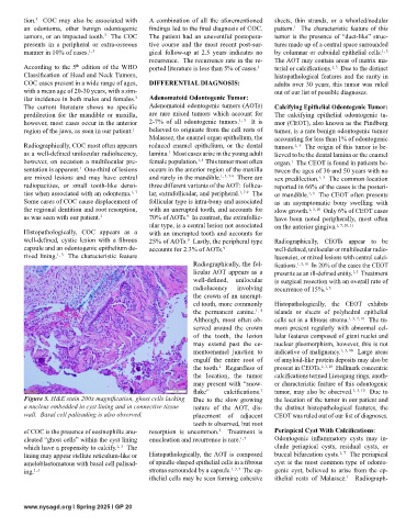
Figure 5. H&E stain 200x magnification, ghost cells lacking Due to the slow growing the location of the tumor in our patient and
a nucleus embedded in cyst lining and in connective tissue nature of the AOT, dis- the distinct histopathological features, the
wall. Basal cell palisading is also observed. placement of adjacent CEOT was ruled out of our list of diagnoses.
teeth is observed, but root
of COC is the presence of eosinophilic anu- resorption is uncommon. Treatment is Periapical Cyst With Calcifications:
3
cleated “ghost cells” within the cyst lining enucleation and recurrence is rare. 1, 7 Odontogenic inflammatory cysts may in-
which have a propensity to calcify. The clude periapical cysts, residual cysts, or
1, 3
1, 7
lining may appear stellate reticulum-like or Histopathologically, the AOT is composed buccal bifurcation cysts. The periapical
ameloblastomatous with basal cell palisad- of spindle-shaped epithelial cells in a fibrous cyst is the most common type of odonto-
ing. 1, 3 stroma surrounded by a capsule. 1, 3, 7 The ep- genic cyst, believed to arise from the ep-
ithelial cells may be seen forming cohesive ithelial rests of Malassez. Radiograph-
1
www.nysagd.org l Spring 2025 l GP 20