
Page 112 - Libro 2
P. 112
92
PART 2 — CEREBROVASCULAR
Originally introduced by Rune Aaslid in 1982 and
applied to patients with vasospasm secondary to sub- A2
ACoA
arachnoid hemorrhage (SAH), transcranial Doppler
(TCD) and transcranial duplex imaging (TCDI) pro-
vide diagnostic information in patients with a variety CS C2 ACA of cerebrovascular diseases.1 This ultrasound tech-
nology complements the neuroimaging techniques
of computed tomography with contrast (CTA), mag-
netic resonance imaging with contrast (MRA), and M2
cerebral angiography by providing physiological data M1 PCoA in real time that can be repeated, which is a valuable ICA
tool when considering the complex dynamics of
cerebral blood flow.
A2
A1 OA
C2 CS
OA A1 C3
C3
M2 M1
ICA P2 PCA
MCA
C1
BA
MCA
ANATOMY
TCD examinations directly study the intracranial conducting arteries that lie at the base of the brain, including the arterial anatomists called the circle of Willis and the major anterior and posterior arteries that supply the circle. To put things in perspective, it is useful to understand that these are small tar- gets; the center of the circle of Willis is about the size of a thumbnail and, on average, the diameter of the basal cerebral arteries range from approximately 2 to 4 mm.2,3
Most cerebral arteries have a numerical classifi- cation system that describes each arterial segment by name and number with the number referring to either the anatomical course or a branch point. Vari- ations in the circle of Willis are frequent in 18% to 54% of individuals and result from anomalies in ves- sel caliber, course, and origin of branches.4,5
The anterior circulation is formed by the intra- cranial continuation of the internal carotid artery (ICA), which first becomes accessible by TCD exam in the cavernous portion, which is usually referred to as the carotid “siphon” because of its tortuous course (Fig. 7-1). The siphon is broken down into three segments: the parasellar (C4), the genu (C3), and the supraclinoid (C2). The ICA pierces the dura, then enters the subarachnoid space, and terminates (C1) by dividing into the middle cerebral (MCA) and anterior cerebral (ACA) arteries. Significant branch- es to a TCD study that arise from the distal ICA are the ophthalmic artery (OA) and posterior communi- cating arteries (PCOAs).
The MCA branches and courses laterally from the ICA as the main trunk or M1 segment and bi- furcates or trifurcates into M2 branches that quickly angle upward into the insular area. There is very little asymmetry between the left and right middle cerebral arteries. The ACA (A1 or precommunicating segment) begins and courses medially from the ICA for a short distance before passing forward as the
PCA P2 P1
VA
P1
VA
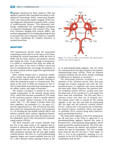
Figure 7-1 Circle of Willis and branches with abbreviations used for each arterial segment.
A2 or postcommunicating segment. The two ACAs are connected above the optic chiasm by the anterior communicating artery (ACOA). There are frequent variations between the two ACAs, mainly consisting of differences in diameter or curvature.4–6
The intracranial posterior circulation is a con- tinuation of the vertebral arteries (VAs) that pass through the foramen magnum and enter the sub- arachnoid space (V4 segment) and course beneath the brain stem. Major branches, the posterior infe- rior cerebellar arteries (PICAs), usually arise from the distal part of the vertebral (V4) and supply the brain stem and cerebellum. The VAs may have a tortuous course and often are of unequal size.6 The basilar artery (BA) is created by the joining together of the two VAs and ends by terminating into the right and left posterior cerebral arteries (PCAs). It gives rise to two paired sets of branches, the anterior inferior cerebellar (AICA) and superi- or cerebellar (SCA) arteries along with numerous, small penetrating branches.7
The initial short segment of the PCA arising from the BA and prior to the PCOA is called the P1 or precommunicating segment. Beyond this point it becomes the P2, postcommunicating segment, which winds around the cerebral peduncle. The PCOA ana- tomically connects the anterior and posterior circu- lations but may be hypoplastic. The PCA normally arises from the BA but can have a “fetal origin” in 18% to 27% of the population, meaning it is de- pendent on the ICA for flow, either exclusively or in combination with the BA.7
