
Page 115 - Libro 2
P. 115
7 — Intracranial Cerebrovascular Examination
95
There are a wide range of normal values that vary primarily due to age, gender, cardiac effects, and by other intrinsic and extrinsic physiologic variables. Waveforms are evaluated for both quantitative and qualitative characteristics and with knowledge of physiological variables that can significantly influ- ence flow findings.
Audio Signals
Doppler signals are audible as well as visible in the spectral waveform. With the increased sophistica- tion of ultrasound instrumentation, there has been a trend toward diminishing the importance of hear- ing Doppler sounds. The human ear and brain are exquisitely designed to perceive subtle audio nu- ances and, when used for vascular studies, provide a feedback loop that informs the hand how to move to acquire a stronger signal and to increase gain to visualize the low amplitude, high velocity (pitch) signals that are otherwise drowned out by the high amplitude, low velocity turbulent signals. Develop- ing good listening skills is especially important dur- ing nonimaging TCD and drives the acquisition of good signals but should not be minimized during TCDI examinations.
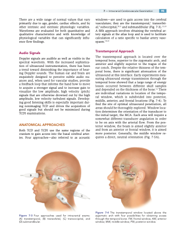
ANATOMICAL APPROACHES
Both TCD and TCDI use the same regions of the cranium to gain access into the basal cerebral arter- ies. Four approaches—also referred to as acoustic
windows—are used to gain access into the cerebral vasculature; they are the transtemporal,1 transorbit- al,9 suboccipital,10,11 and submandibular (Fig. 7-3).12,13 A fifth approach involves obtaining the vertebral ar- tery signals at the atlas loop and is used to facilitate calculation of a ratio specific to basilar artery vaso- spasm.14,15
Transtemporal Approach
The transtemporal approach is located over the temporal bone, superior to the zygomatic arch, and anterior and slightly superior to the tragus of the ear conch. Despite the relative thinness of the tem- poral bone, there is significant attenuation of the ultrasound at this interface. Early experiments mea- suring ultrasound energy transmission through the temporal bone showed that a large range of energy losses occurred between different skull samples and depended on the thickness of the bone.16 There are individual variations in location of the tempo- ral window, which is subdivided into posterior, middle, anterior, and frontal locations (Fig. 7-4). To find the site of optimal ultrasound penetration, all areas should be thoroughly explored. Window loca- tion determines the orientation of the transducer to the initial target, the MCA. Each area will require a somewhat different transducer angulation in order to be on axis with the arterial flow. From the pos- terior window, the beam is aimed slightly anterior and from an anterior or frontal window, it is aimed more posterior. Generally, the middle window re- quires a direct, neutral orientation (Fig. 7-5A).
FW
AW MW PW
Figure 7-4 The transtemporal window located above the zygomatic arch with four possibilities for obtaining access through the temporal bone. FW, frontal window; AW, anterior window; MW, middle window; PW, posterior window.
B
Figure 7-3 Four approaches used for intracranial exams: (A) transtemporal, (B) transorbital, (C) transoccipital, and (D) submandibular.
A
D
C
