
Page 130 - Libro 2
P. 130
110
PART 2 — CEREBROVASCULAR
Submandibular Approach
The retromandibular extradural ICA is routinely ob- tained in patients with abnormalities, requiring cal- culation of the Lindegaard ratio including SAH, head trauma, intracranial stenosis, and arteriovenous mal- formations. This ratio, defined as MCA/SM-ICA, is important for differentiating vasospasm and stenosis of the MCA from hyperdynamic flow.13,21 If the patient has 50% extracranial stenosis, the ratio calculation may be invalid and should not be used. Locating the ICA using this technique is also useful in determining distal arterial narrowing, which is often associated with carotid dissection or fibromuscular dysplasia.
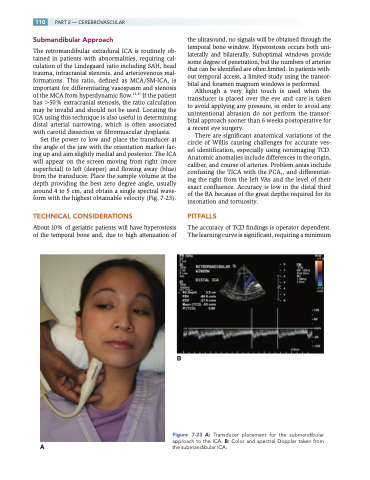
Set the power to low and place the transducer at the angle of the jaw with the orientation marker fac- ing up and aim slightly medial and posterior. The ICA will appear on the screen moving from right (more superficial) to left (deeper) and flowing away (blue) from the transducer. Place the sample volume at the depth providing the best zero degree angle, usually around 4 to 5 cm, and obtain a single spectral wave- form with the highest obtainable velocity (Fig. 7-23).
TECHNICAL CONSIDERATIONS
About 10% of geriatric patients will have hyperostosis of the temporal bone and, due to high attenuation of
Figure 7-23 A: Transducer placement for the submandibular
approach to the ICA. B: Color and spectral Doppler taken from A the submandibular ICA.
the ultrasound, no signals will be obtained through the temporal bone window. Hyperostosis occurs both uni- laterally and bilaterally. Suboptimal windows provide some degree of penetration, but the numbers of arteries that can be identified are often limited. In patients with- out temporal access, a limited study using the transor- bital and foramen magnum windows is performed.
Although a very light touch is used when the transducer is placed over the eye and care is taken to avoid applying any pressure, in order to avoid any unintentional abrasion do not perform the transor- bital approach sooner than 6 weeks postoperative for a recent eye surgery.
There are significant anatomical variations of the circle of Willis causing challenges for accurate ves- sel identification, especially using nonimaging TCD. Anatomic anomalies include differences in the origin, caliber, and course of arteries. Problem areas include confusing the TICA with the PCA1, and differentiat- ing the right from the left VAs and the level of their exact confluence. Accuracy is low in the distal third of the BA because of the great depths required for its insonation and tortuosity.
PITFALLS
The accuracy of TCD findings is operator dependent. The learning curve is significant, requiring a minimum
B
