
Page 162 - Libro 2
P. 162
142
PART 3 — PERIPHERAL ARTERIAL
examination. There are classic symptoms that may indicate the presence of arterial insufficiency or isch- emia including intermittent claudication, rest pain, nonhealing ulcers, and gangrene. There can be some subtle changes such as hair loss, nail thickening, and skin changes. Symptoms such as pallor, pulse- lessness, paralysis, paresthesia, and intense pain are indicative of acute arterial ischemia. Peripheral arte- rial aneurysms may also be suspected if upon palpa- tion a mass is detected in the femoral or popliteal regions. Peripheral arteries may also be examined for aneurysmal disease if the patient has a known abdominal aortic aneurysm. Atherosclerosis and an- eurysmal disease are the primary pathologies sus- pected in most individuals. There are less frequently encountered arterial diseases, as well as traumatic and iatrogenic injuries, which may require DA for their diagnosis.
Although not an indication for testing, many of the patients will present with comorbid risk factors. These risk factors include diabetes, hyperlipidemia, hypertension, history of tobacco use, coronary artery disease, and end-stage renal disease. Additional risk factors for arterial disease are obesity, a sedentary lifestyle, heredity, gender, and age.
SONOGRAPHIC EXAMINATION TECHNIQUES
PATIENT PREPARATION
The patient should have the test procedure explained to him or her. The patient should remove all clothing from the waist down, except for undergarments, and be given a patient gown or appropriate drape.
PATIENT POSITIONING
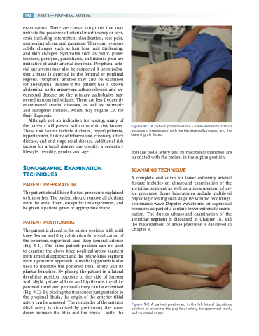
The patient is placed in the supine position with mild knee flexion and thigh abduction for visualization of the common, superficial, and deep femoral arteries (Fig. 9-1). The same patient position can be used to examine the above-knee popliteal artery segment from a medial approach and the below-knee segment from a posterior approach. A medial approach is also used to insonate the posterior tibial artery and its plantar branches. By placing the patient in a lateral decubitus position opposite to the side of interest with slight ipsilateral knee and hip flexure, the tibio- peroneal trunk and peroneal artery can be examined (Fig. 9-2). By placing the transducer just posterior to the proximal fibula, the origin of the anterior tibial artery can be assessed. The remainder of the anterior tibial artery is visualized by positioning the trans- ducer between the tibia and the fibula. Lastly, the
Figure 9-1 A patient positioned for a lower extremity arterial ultrasound examination with the hip externally rotated and the knee slightly flexed.
dorsalis pedis artery and its metatarsal branches are insonated with the patient in the supine position.
SCANNING TECHNIQUE
A complete evaluation for lower extremity arterial disease includes an ultrasound examination of the aortoiliac segment as well as a measurement of an- kle pressures. Some laboratories include multilevel physiologic testing such as pulse volume recordings, continuous-wave Doppler waveforms, or segmental pressures as part of a routine lower extremity exami- nation. The duplex ultrasound examination of the aortoiliac segment is discussed in Chapter 18, and the measurement of ankle pressures is described in Chapter 8.
Figure 9-2 A patient positioned in the left lateral decubitus position to examine the popliteal artery, tibioperoneal trunk, and peroneal artery.
