
Page 164 - Libro 2
P. 164
144
PART 3 — PERIPHERAL ARTERIAL
Figure 9-6 The posterior tibial (PTA) and peroneal arteries arising off the tibioperoneal trunk (TPT).
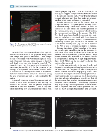
Individual laboratory protocols vary, but typically include documentation of the grayscale image, color flow image, and spectral waveforms. These images should be recorded over every major segment evalu- ated. Proximal, mid, and distal images of the SFA and tibial vessel are also typically recorded. The grayscale image may be documented in sagittal and transverse planes (Fig. 9-7). In the presence of pa- thology, it is important to document the full extent of the disease. If aneurysmal disease is suspected, diameter measurements should be recorded along the area of concern as well as just proximal to this region.
In general, color and power Doppler are used pri- marily to assist with localization and tracking the course of the vessels. Color can provide a rapid as- sessment of the flow dynamics. Color is very use- ful in identifying flow abnormalities associated with
arterial plaque (Fig. 9-8). Color is also helpful in placing the Doppler sample volume at the area of the of the greatest velocity shift. Power Doppler should be used whenever very low flow states are encoun- tered or when vessel occlusion is suspected.
Velocity spectra are used as the primary tool to categorize disease. The peak systolic velocity (PSV) is recorded along all the major vessels. In areas of a stenosis, the PSV should be recorded proximal to the stenosis, at the area of maximum velocity shift in the stenosis, and just distal to the stenosis (Fig. 9-9). The distal waveform should demonstrate the post- stenotic turbulence associated with hemodynami- cally significant stenoses. The PSV at the stenosis is divided by the PSV just proximal to the stenosis to calculate the velocity ratio (Vr). The Vr, in addition to the PSV, is used to estimate the degree of stenosis.
Because the status of the branches of the arter- ies can also add valuable data for the surgeon, vi- sualization of as many tibial and pedal branches as possible including maleolar, plantars, tarsals, deep plantar arteries, and branches of the named vessels is also performed during DA. A high-frequency trans- ducer (15-7 MHz) can be especially useful in this portion of the protocol.
A precise evaluation of arterial size, length, and degree of narrowing as well as plaque characteristics are performed for a single focal lesion or sequential lesions suitable for balloon angioplasty and/or stent placement. It is important for the sonographer or vas- cular technologist to possess as much information from the referring physician as possible so that all the ultrasound data required for patient management decisions can be obtained. The referring physician may be planning a specific procedure or an interven- tion at a specific level and require pertinent data to plan the most appropriate procedure and approach.
AB
Figure 9-7 A: A sagittal scan of an artery with atherosclerotic plaque. B: A transverse view of the same plaque (arrow).
