
Page 166 - Libro 2
P. 166
146
PART 3 — PERIPHERAL ARTERIAL
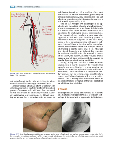
Figure 9-10 An arterial map drawing of a patient with multiple failed PTFE bypasses.
not routinely used for the entire arterial tree; therefore, eccentric arterial lesions may go undetected by CA.
Another unique advantage of DA as compared to other imaging tools is its ability to identify the softest portion of the vessel wall, which can then be marked on the skin before the intended procedure. Exten- sive calcification in a vessel makes for difficult sutur- ing, so an area that is compliant with no plaque or
calcification is preferred. Skin marking of the most suitable site for outflow anastomosis, particularly for infrapopliteal segments, may limit incision size and eliminate extensive arterial dissection in search of a soft arterial segment (Fig. 9-11).
One of the strongest DA advantages is its ap- plication in the setting of acute arterial ischemia.22 Current management of acute lower limb ischemia has evolved from simple embolectomies under local anesthesia to challenging arterial reconstructions. This dramatic change involves a more aggressive approach at limb salvage in the elderly patient by well-trained vascular surgeons. On the other hand, many of these patients presenting with acutely isch- emic limbs will have underlying multisegmental oc- clusive arterial disease rather than a simple embolus obstructing a healthy vessel (Fig. 9-12). Although the clinical diagnosis of an ischemic leg can often be made without difficulties, the anatomical pattern of the inflow, the outflow, and the occluded arterial segment may at times be impossible to ascertain by standard preoperative imaging modalities.
Finally, during the course of a lower extremity examination, it may be necessary to evaluate other vascular segments. Routinely, venous mapping can also be performed during DA to identify usable veins for harvest. The examination of the subclavian–axil- lary segment may be performed as a possible inflow source for debilitated patients with severe aortoiliac disease. This is accomplished without the risk of an additional thoracic aortogram or the time needed for an additional thoracic MRA.
PITFALLS
Investigators have clearly demonstrated the feasibility and multiple advantages of DA and, as with any tech- nology, it is important to appreciate its limitations.
Figure 9-11 Left: Distal posterior tibial artery segment and a large inflow branch are marked preoperatively on the skin. Right: Intraoperative completion angiogram of the newly created vein bypass demonstrated a normal distal anastomosis to the distal posterior tibial artery and preserved large collateral branch on the same patient.
