
Page 168 - Libro 2
P. 168
148
PART 3 — PERIPHERAL ARTERIAL
TRAINING METHODS
To successfully achieve these results is to establish the training method for sonographers/technologists and surgeons interpreting the results and planning revascularization strategy. In an attempt to develop a training period at the author’s institution, the first 25 exams completed by any new sonographer or vas- cular technologist are prospectively confirmed with CA or repeated DA examination by an established staff technical member. In an effort to facilitate the advancement of the DA protocol, every completion angiogram is reviewed with the staff member who performed the exam, as are the iliac angioplasties. The characteristics of the proximal and distal arter- ies, vein conduit, or tibial vein (in the case when an adjunctive arteriovenous fistula is performed) are discussed and any discrepancies are reviewed as a quality assurance measure. The technical staff visit the operating room to witness the intraopera- tive findings firsthand. In this manner, the constant feedback becomes the cornerstone for the continual improvement in the quality of the DA exams.
DIAGNOSIS
GRAYSCALE FINDINGS
Normal arterial walls appear smooth and uniform. As the atherosclerotic disease progresses, the vessel walls will thicken. Calcification may be present and will produce acoustic shadowing, limiting complete evaluation of the vessel. Vessel wall thickness and de- gree of calcification are reported to aid in the choice of anastomosis sites (Fig. 9-14). As stated earlier, a sur- geon will want to avoid areas of heavy calcification because it is difficult to pass sutures through heav- ily calcified vessels. Plaque can be seen encroach- ing on the vessel lumen. Most plaque will appear
Figure 9-14 Color Doppler image of a distal posterior tibial artery with segmental heavy calcifications (arrows) creating shadows obscuring arterial lumen.
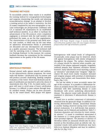
Figure 9-15 Power Doppler image of severely diseased, behind-the-knee popliteal artery with very irregular ulcerated plaque surface with high embolization potential.
heterogeneous with mixed levels of echogenicity. Occasionally, some plaque may be observed, which will appear homogeneous with similar echogenicity throughout the plaque. The surface characteristics of a plaque should be reported when possible, par- ticularly if the plaque appeared irregular. Irregularly surfaced plaques may represent ulcerative lesions; however, many laboratories avoid reporting a plaque as “ulcerative” and simply state “irregular.” High res- olution DA much more clearly visualizes ulcerated and irregular plaques with potential of embolization plaques, their surface, and characteristic flow distur- bance (Fig. 9-15).
DA has the ability to more accurately assess the age of the occlusion. It is possible to differentiate be- tween an isolated chronic SFA occlusion and an acute embolism with little underlying disease or acute thrombosis with severe underlying atherosclerotic disease. The adjacent vessel walls should be closely examined to determine if atherosclerotic disease is present.
Vessel size is another component of information obtained from the grayscale image. In addition to de- tecting atherosclerotic disease, aneurysmal disease is another finding observed during lower extremity arterial evaluations. It is well-known that aneurys- mal disease can be bilateral and multilevel. Table 9-1 illustrates diameters and velocities for lower extrem- ity arteries.25 A vessel is considered aneurysmal if the diameter is 1.5 times greater than the adjacent, more proximal segment. The presence or absence of thrombus within an aneurysm should also be docu- mented as this thrombus poses an embolic risk. An- eurysmal vessels with partial thrombosis may have little-to-no luminal dilatation and may be undetect- able by CA (Fig. 9-16).26
