
Page 167 - Libro 2
P. 167
9 — Duplex Ultrasound of Lower Extremity Arteries
147
Figure 9-12 Color Doppler image of an occluded SFA with acute thrombus overlying severe chronic arterial disease.
Overall, the most common problem with DA has been with calcification. However, some techniques can be used to obtain the necessary information even within severely calcified vessel such as using multiple in- sonation planes, increasing color and Power Doppler gain, sensitivity and persistence, or using SonoCT imaging mode.
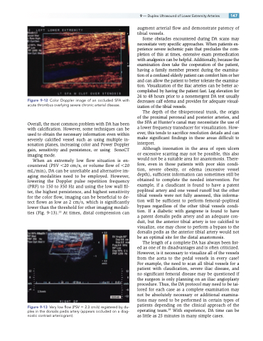
When an extremely low flow situation is en- countered (PSV 20 cm/s, or volume flow of 20 mL/min), DA can be unreliable and alternative im- aging modalities need to be employed. However, lowering the Doppler pulse repetition frequency (PRF) to 150 to 350 Hz and using the low wall fil- ter, the highest persistence, and highest sensitivity for the color flow, imaging can be beneficial to de- tect flows as low as 2 cm/s, which is significantly lower than the threshold for other imaging modali- ties (Fig. 9-13).23 At times, distal compression can
Figure 9-13 Very low flow (PSV 2.3 cm/s) registered by du- plex in the dorsalis pedis artery (appears occluded on a diag- nostic contrast arteriogram).
augment arterial flow and demonstrate patency of tibial vessels.
Some obstacles encountered during DA scans may necessitate very specific approaches. When patients ex- perience severe ischemic pain that precludes the com- pletion of this at times, extensive exam premedication with analgesics can be helpful. Additionally, because the examination does take the cooperation of the patient, having a family member present during the examina- tion of a confused elderly patient can comfort him or her and can allow the patient to better tolerate the examina- tion. Visualization of the iliac arteries can be better ac- complished by having the patient fast. Leg elevation for 24 to 48 hours prior to a nonemergent DA test usually decreases calf edema and provides for adequate visual- ization of the tibial vessels.
The depth of the tibioperoneal trunk, the origin of the proximal peroneal and posterior arteries, and the SFA at Hunter’s canal may necessitate the use of a lower frequency transducer for visualization. How- ever, this tends to sacrifice resolution details and can make significant findings in these areas difficult to interpret.
Although insonation in the area of open ulcers or excessive scarring may not be possible, this also would not be a suitable area for anastomosis. There- fore, even in those patients with poor skin condi- tion, severe obesity, or edema (excessive vessel depth), sufficient information can sometimes still be obtained to complete the needed intervention. For example, if a claudicant is found to have a patent popliteal artery and one vessel runoff but the other tibial vessels were not fully assessed, this informa- tion will be sufficient to perform femoral–popliteal bypass regardless of the other tibial vessels condi- tion. If a diabetic with gangrene is found to have a patent dorsalis pedis artery and an adequate con- duit, but the anterior tibial artery is too calcified to visualize, one may chose to perform a bypass to the dorsalis pedis as the anterior tibial artery would not be an optimal site for the distal anastomosis.
The length of a complete DA has always been list- ed as one of its disadvantages and is often criticized. However, is it necessary to visualize all of the vessels from the aorta to the pedal vessels in every case? For example, the need to scan all tibial vessels for a patient with claudication, severe iliac disease, and no significant femoral disease may be questioned if the surgeon is only planning on an iliac angioplasty procedure. Thus, the DA protocol may need to be tai- lored for each case as a complete examination may not be absolutely necessary or additional examina- tions may need to be performed in certain types of patients depending on the clinical approach of the operating team.24 With experience, DA time can be as little as 25 minutes in many simple cases.
