
Page 170 - Libro 2
P. 170
150
PART 3 — PERIPHERAL ARTERIAL
Figure 9-18 A normal multiphasic waveform taken from SFA.
which results in a multiphasic waveform (Fig. 9-18). There is a sharp upstroke to peak systole, a rapid de- celeration, a reflected wave displayed as retrograde flow below the baseline, and often a small brief wave of antegrade flow in diastole. In situations where the peripheral resistance is lowered, constant forward flow through diastole may be observed. In the cases of a distal arteriovenous fistula, trauma, or cellulitus in the postexercise patient, antegrade flow will be observed throughout diastole (Fig. 9-19). However, in these cases, a normal sharp upstroke to peak systole will be maintained. In the event of significant arterial disease or an occlusion, the vessels distal to this disease will display a low resistance signal with antegrade flow through diastole, but there will be a delayed rise to peak systole (Fig. 9-20). Lastly, when scanning vessels proximal to an occlusion or near occlusion, the spectral waveforms can display a very high resistance pattern
Figure 9-19 A waveform displaying a normal systolic upstroke with constant forward flow through diastole. This can be ob- served immediately after exercise or in the presence of a distal arteriovenous fistula, trauma, or cellulitus.
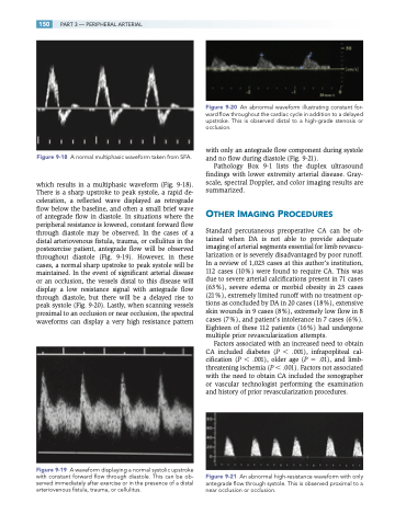
Figure 9-20 An abnormal waveform illustrating constant for- ward flow throughout the cardiac cycle in addition to a delayed upstroke. This is observed distal to a high-grade stenosis or occlusion.
with only an antegrade flow component during systole and no flow during diastole (Fig. 9-21).
Pathology Box 9-1 lists the duplex ultrasound findings with lower extremity arterial disease. Gray- scale, spectral Doppler, and color imaging results are summarized.
OTHER IMAGING PROCEDURES
Standard percutaneous preoperative CA can be ob- tained when DA is not able to provide adequate imaging of arterial segments essential for limb revascu- larization or is severely disadvantaged by poor runoff. In a review of 1,023 cases at this author’s institution, 112 cases (10%) were found to require CA. This was due to severe arterial calcifications present in 71 cases (63%), severe edema or morbid obesity in 23 cases (21%), extremely limited runoff with no treatment op- tions as concluded by DA in 20 cases (18%), extensive skin wounds in 9 cases (8%), extremely low flow in 8 cases (7%), and patient’s intolerance in 7 cases (6%). Eighteen of these 112 patients (16%) had undergone multiple prior revascularization attempts.
Factors associated with an increased need to obtain CA included diabetes (P .001), infrapopliteal cal- cification (P .001), older age (P .01), and limb- threatening ischemia (P .001). Factors not associated with the need to obtain CA included the sonographer or vascular technologist performing the examination and history of prior revascularization procedures.
Figure 9-21 An abnormal high-resistance waveform with only antegrade flow through systole. This is observed proximal to a near occlusion or occlusion.
