
Page 18 - Libro 2
P. 18
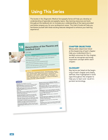
                                  The books in the Diagnostic Medical Sonography Series will help you develop an understanding of specialty sonography topics. Key learning resources and tools throughout the textbook aim to increase your understanding of the topics provided and better prepare you for your professional career. This User’s Guide will help you familiarize yourself with these exciting features designed to enhance your learning experience.
CHAPTER OBJECTIVES
Measurable objectives listed
at the beginning of each chapter help you understand the in- tended outcomes for the chapter, as well as recognize and study important concept within each chapter.
GLOSSARY
Key terms are listed at the begin- ning of each chapter and clearly defined, then highlighted in bold type throughout the chapter to help you to learn and recall im- portant terminology.
              xvi
Using This Series
  425 GLOSSARY
18 Abnormalities of the Placenta and Umbilical Cord
Aneurysm Focal dilatation of an artery
Bilobed placenta Placenta where the lobes are nearly
Marginal insertion (a.k.a. battledore placenta)
equal in size and the cord inserts into the chorionic bridge of tissue that connects the two lobes
Body stalk anomaly Fatal condition associated with multiple congenital anomalies and absence of the um- bilical cord
Occurs when the umbilical cord inserts at the placental margin instead of centrally
Mickey Mouse sign Term used to describe the cross-section of the three-vessel umbilical cord or the portal triad (portal vein, hepatic artery, common bile duct)
Breus’ mole Very rare condition where there is massive subchorionic thrombosis of the placenta secondary to extreme venous obstruction
Extrachorial placenta Attachment of the placental membranes to the fetal surface of the placenta rather than to the underlying villous placental margin
Omphalocele Central anterior abdominal wall defect of the umbilicus where abdominal organs are contained by a covering membrane consisting of peritoneum, Wharton’s jelly, and amnion
Placentomegaly Term that refers to a thickened placenta
Synechia (Asherman’s syndrome) Linear, extra amni- otic tissue that projects into the amniotic cavity with no restriction of fetal movement
Thrombosis Intraplacental area of hemorrhage
and clot
True knot Result of the fetus actually passing through a loop or loops of umbilical cord creating one or more knots in the cord
False knot Bending, twisting, and bulging of the umbil- ical cord vessels mimicking a knot in the umbilical cord Gastroschisis Periumbilical abdominal wall defect, typically to the right of normal cord insertion, that al- lows for free-floating bowel in the amniotic fluid Limb–body wall complex Condition characterized by
multiple complex fetal anomalies and a short umbilical cord
Lisa M. Allen
OBJECTIVES
Recognize the sonographic appearance of placental and umbilical cord anomalies Discuss developmental variations in placental size, shape, and configuration Identify placenta previa classifications
Explain placental abruption and the associated risk factors
List placenta accreta classifications and known risk factors
Name the various abnormalities of umbilical cord insertion into the placenta Describe cystic and solid masses of the umbilical cord
KEY TERMS
succenturiate lobe | circummarginate placenta | circumvallate placenta | placenta previa | placental abruption | placenta accreta spectrum | chorioangioma | amniotic band syndrome | uterine synechiae | marginal insertion | battledore placenta | velamentous insertion | true knot | false knot | nuchal cord | cord prolapse | vasa previa | single umbilical artery | cord entanglement | umbilical cord hemangioma | umbilical cord coiling | umbilical coiling index
425
10/22/11
3:30 AM
